

Progression of Unilateral Hearing Loss in Children With and Without Ipsilateral Cochlear Nerve Canal Stenosis: A Hazard Analysis
- PMID: 28538470
- PMCID: PMC5639713
- DOI: 10.1097/MAO.0000000000001452
Progression of Unilateral Hearing Loss in Children With and Without Ipsilateral Cochlear Nerve Canal Stenosis: A Hazard Analysis
Abstract
Objective: To investigate the risk of hearing loss progression in each ear among children with unilateral hearing loss associated with ipsilateral bony cochlear nerve canal (BCNC) stenosis.
Setting: Tertiary pediatric referral center.
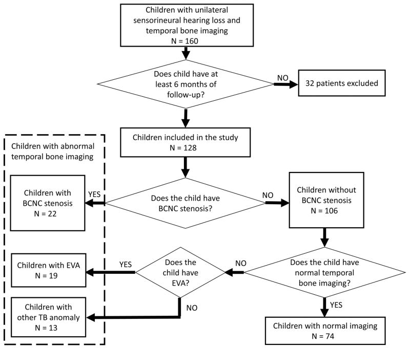
Patients: Children diagnosed with unilateral hearing loss who had undergone temporal bone computed tomography imaging and had at least 6 months of follow-up audiometric testing were identified from a prospective audiological database.
Interventions: Two pediatric radiologists blinded to affected ear evaluated imaging for temporal bone anomalies and measured bony cochlear canal width independently. All available audiograms were reviewed, and air conduction thresholds were documented.
Main outcome measure: Progression of hearing loss was defined by a 10 dB increase in air conduction pure-tone average.
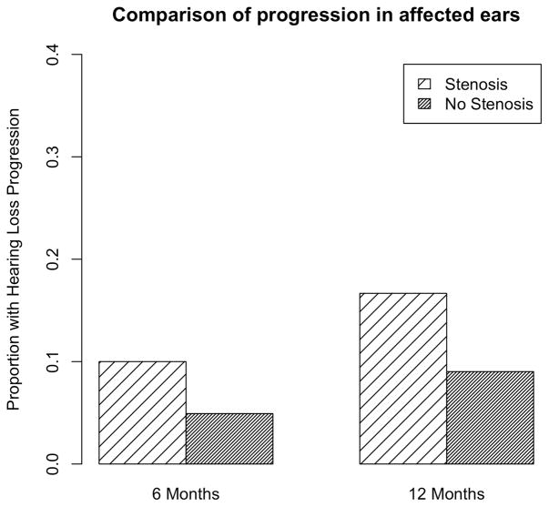
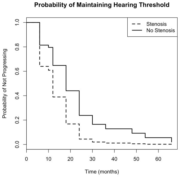
Results: One hundred twenty eight children met inclusion criteria. Of these, 54 (42%) had a temporal bone anomaly, and 22 (17%) had ipsilateral BCNC stenosis. At 12 months, rates of progression in the ipsilateral ear were as follows: 12% among those without a temporal bone anomaly, 13% among those with a temporal bone anomaly, and 17% among those with BCNC stenosis. Children with BCNC stenosis had a significantly greater risk of progression in their ipsilateral ear compared with children with no stenosis: hazard ratio (HR) 2.17, 95% confidence interval (CI) (1.01, 4.66), p value 0.046. When we compared children with BCNC stenosis to those with normal temporal bone imaging, we found that the children with stenosis had nearly two times greater risk estimate for progression, but this difference did not reach significance, HR 1.9, CI (0.8, 4.3), p = 0.1. No children with BCNC stenosis developed hearing loss in their contralateral year by 12 months of follow-up.
Conclusion: Children with bony cochlear nerve canal stenosis may be at increased risk for progression in their ipsilateral ear. Audiometric and medical follow-up for these children should be considered.
Figures
References
-
- Simons JP, Mandell DL, Arjmand EM. Computed Tomography and Magnetic Resonance Imaging in Pediatric Unilateral and Asymmetric Sensorineural Hearing Loss. Arch Otolaryngology Head and Neck Surgery. 2006;132(2):186–192. - PubMed
-
- Friedman AB, Guillory R, Ramakrishnaiah RH, Frank R, Gluth MB, Richter GT, Dornhoffer JL. Risk analysis of unilateral severe-to-profound sensorineural hearing loss in children. Int J Pediatr Otorhinolaryngol. 2013;77(7):1128–31. - PubMed
-
- Adunka OF, Jewells V, Buchman CA. Value of Computed Tomography in the Evaluation of Children with Cochlear Nerve Deficiency. Otology and Neurotology. 2007;28:597–604. - PubMed
-
- DeMarcantonio M, Choo DI. Radiographic evaluation of children with hearing loss. Otolaryngol Clin North Am. 2015;48(6):913–32. - PubMed
-
- Song J, Choi HG, Oh SH, Chang SO, Kim CS, Lee JH. Unilateral Sensorineural Hearing Loss in Children: The Importance of Temporal Bone Computed Tomography and Audiometric Follow-Up. Otology and Neurotology. 2009;30:604–608. - PubMed
MeSH terms
Grants and funding
LinkOut - more resources
Full Text Sources
Other Literature Sources
Miscellaneous
