

Cutaneous angiosarcoma: report of three different and typical cases admitted in a unique dermatology clinic
- PMID: 28538886
- PMCID: PMC5429112
- DOI: 10.1590/abd1806-4841.20175326
Cutaneous angiosarcoma: report of three different and typical cases admitted in a unique dermatology clinic
Abstract
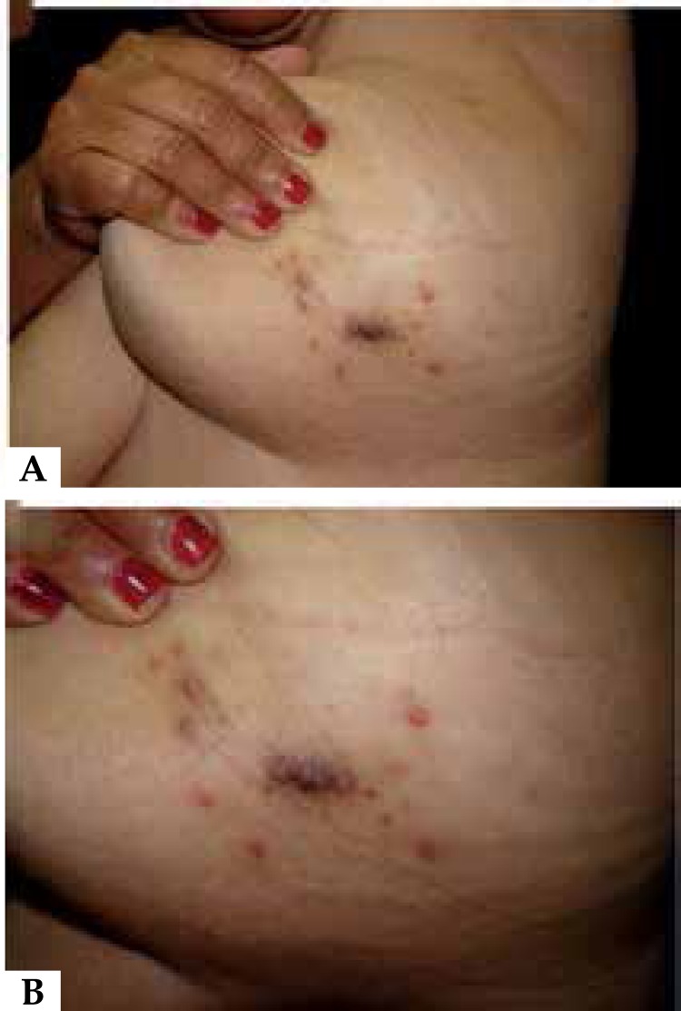
Angiosarcoma is a rare and aggressive tumor with high rates of metastasis and relapse. It shows a particular predilection for the skin and superficial soft tissues. We report three distinct and typical cases of angiosarcoma that were diagnosed in a single dermatology clinic over the course of less than a year: i) Angiosarcoma in lower limb affected by chronic lymphedema, featuring Stewart-Treves syndrome; ii) a case of the most common type of angiosarcoma loated in the scalp and face of elderly man and; iii) a skin Angiosarcoma in previously irradiated breast. All lesions presented characteristic histopathological findings: irregular vascular proliferation that dissects the collagen bundles with atypical endothelial nuclei projection toward the lumen.
Conflict of interest statement
Conflict of interest: none.
Figures


References
-
- Young RJ, Brown NJ, Reed MW, Hughes D, Woll PJ. Angiosarcoma. Lancet Oncol. 2010;11:983–991. - PubMed
-
- Penel N, Marréaud S, Robin YM, Hohenberger P. Angiosarcoma: State of the art and perspectives. Crit Rev Oncol Hematol. 2011;80:257–263. - PubMed
-
- Albores-Saavedra J, Schwartz AM, Henson DE, Kostun L, Hart A, Angeles-Albores D, et al. Cutaneous angiosarcoma. Analysis of 434 cases from the Surveillance, Epidemiology, and End Results Program, 1973-2007. Ann Diagn Pathol. 2011;15:93–97. - PubMed
Publication types
MeSH terms
Supplementary concepts
LinkOut - more resources
Full Text Sources
Other Literature Sources
Medical