. 2017 Jul 1;199(1):186-203.
doi: 10.4049/jimmunol.1700145. Epub 2017 May 24.

Bolstering the Number and Function of HSV-1-Specific CD8+ Effector Memory T Cells and Tissue-Resident Memory T Cells in Latently Infected Trigeminal Ganglia Reduces Recurrent Ocular Herpes Infection and Disease

Affiliations

Bolstering the Number and Function of HSV-1-Specific CD8+ Effector Memory T Cells and Tissue-Resident Memory T Cells in Latently Infected Trigeminal Ganglia Reduces Recurrent Ocular Herpes Infection and Disease

Arif A Khan et al. J Immunol. .

Abstract

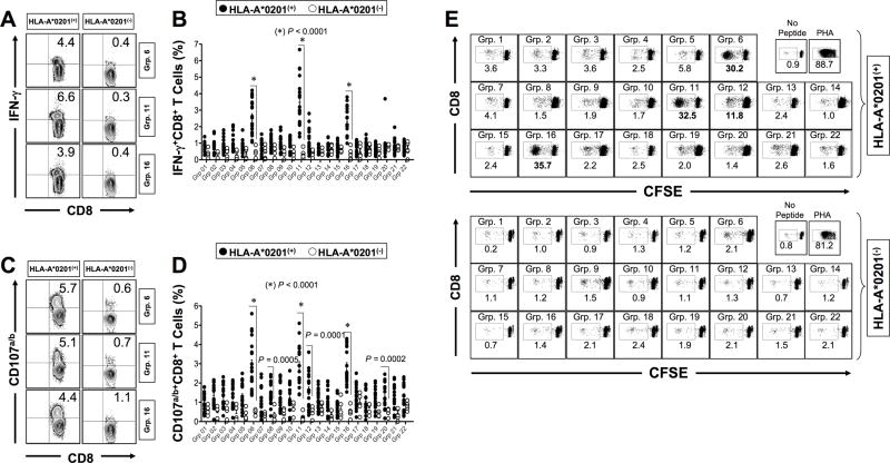
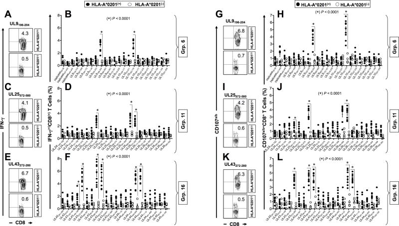
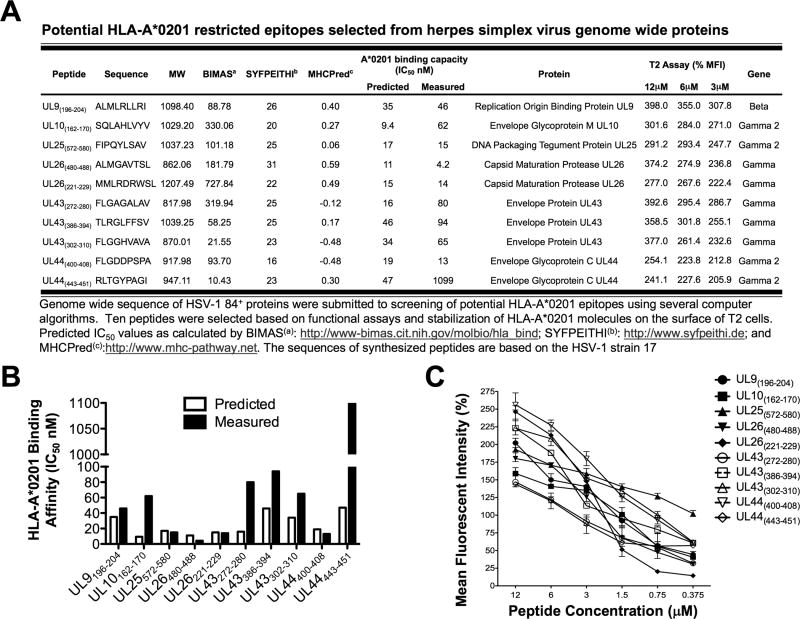
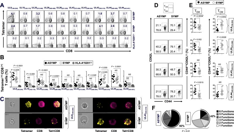
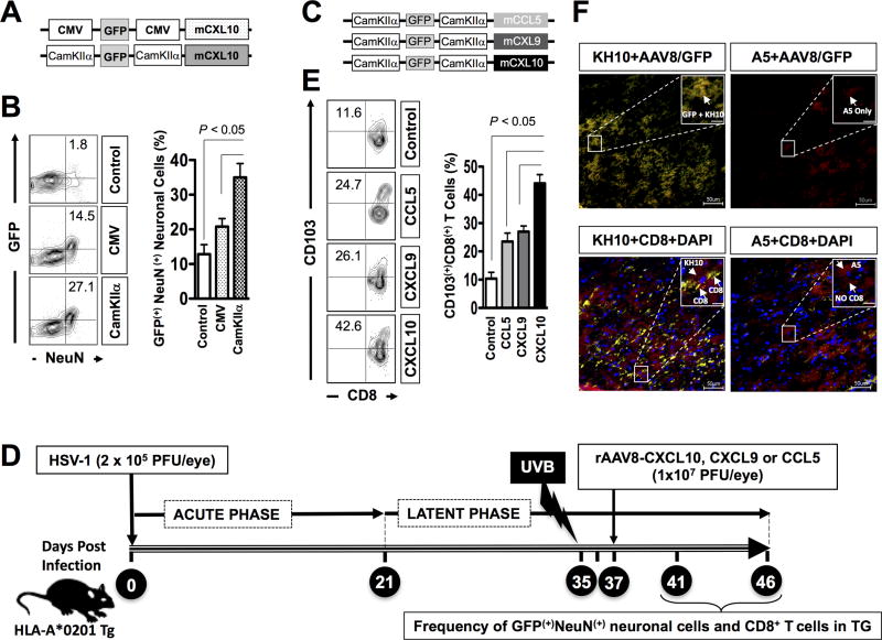
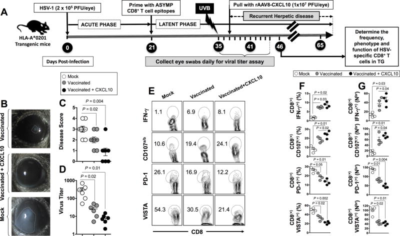
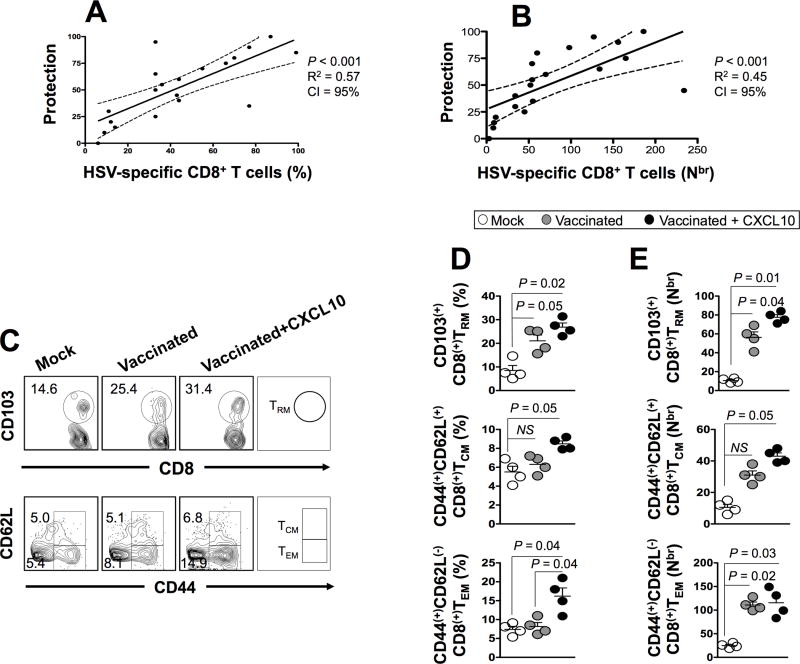
HSV type 1 (HSV-1) is a prevalent human pathogen that infects >3.72 billion individuals worldwide and can cause potentially blinding recurrent corneal herpetic disease. HSV-1 establishes latency within sensory neurons of trigeminal ganglia (TG), and TG-resident CD8+ T cells play a critical role in preventing its reactivation. The repertoire, phenotype, and function of protective CD8+ T cells are unknown. Bolstering the apparent feeble numbers of CD8+ T cells in TG remains a challenge for immunotherapeutic strategies. In this study, a comprehensive panel of 467 HLA-A*0201-restricted CD8+ T cell epitopes was predicted from the entire HSV-1 genome. CD8+ T cell responses to these genome-wide epitopes were compared in HSV-1-seropositive symptomatic individuals (with a history of numerous episodes of recurrent herpetic disease) and asymptomatic (ASYMP) individuals (who are infected but never experienced any recurrent herpetic disease). Frequent polyfunctional HSV-specific IFN-γ+CD107a/b+CD44highCD62LlowCD8+ effector memory T cells were detected in ASYMP individuals and were primarily directed against three "ASYMP" epitopes. In contrast, symptomatic individuals have more monofunctional CD44highCD62LhighCD8+ central memory T cells. Furthermore, therapeutic immunization with an innovative prime/pull vaccine, based on priming with multiple ASYMP epitopes (prime) and neurotropic TG delivery of the T cell-attracting chemokine CXCL10 (pull), boosted the number and function of CD44highCD62LlowCD8+ effector memory T cells and CD103highCD8+ tissue-resident T cells in TG of latently infected HLA-A*0201-transgenic mice and reduced recurrent ocular herpes following UV-B-induced reactivation. These findings have profound implications in the development of T cell-based immunotherapeutic strategies to treat blinding recurrent herpes infection and disease.


Conflict of interest statement

Conflict of interest: The authors have declared that no conflict of interest exists

Figures

Figure 1
Figure 1. Frequency of IFN-γ-producing CD8+ T cells specific to genome wide-derived epitopes in HLA-A*0201(+) HSV-seropositive individuals
PBMCs (~10 × 106) derived from 20 HLA-A*0201(+) individuals vs. 8 HLA-A*0201(−) HSV-seropositive individuals were stimulated in vitro for 72 hrs with each of 22 groups of equimolar peptides. IFN-γ and CD107a/b were then measured by intracellular staining, as outlined in Materials and Methods. (A) Representative contour plots of percentage of IFN-γ+CD8+ T cells specific to groups of HSV-1 peptides 6, 11 and 16 detected in HLA-A*0201(+) (left panels) and HLA-A*0201(−) (right panels) individuals. (B) Average frequencies of IFN-γ+CD8+ T cells specific to 22 groups of HSV-1 peptides detected in PBMCs from HLA-A*0201(+) (closed circle) and HLA-A*0201(−) (open circle) individuals. (C) Representative contour plots of expression of CD107a/b by CD8+ T cells specific to groups of HSV-1 peptides 6, 11 and 16 detected in HLA-A*0201(+) (left panel) and HLA-A*0201(−) (right panel) individuals. (D) Average frequencies of CD107a/b(+)CD8+ T cells detected in HLA-A*0201(+) (closed circle) and HLA-A*0201(−) (open circle) individuals in response to 22 groups of HSV-1 peptides. (E) Representative dot plots of percentage of dividing CFSE(+)CD8+ T cells from HLA-A*0201(+) (top three rows) and HLA-A*0201(−) individuals (lower three rows) following a 6-days in vitro stimulation with each of the 22 groups of HSV-1 peptides. The results are representative of 2 independent experiments. The nominal P values indicated on panels (B) and (D) show statistical significance between HLA-A*0201(+) and HLA-A*0201(−) individuals. P values are calculated using either the parametric two-sample Student’s t-test or non-parametric Wilcoxon rank sum test, as appropriate. For paired comparisons involving the 22 groups of peptides, we have adjusted for multiple comparisons using the Bonferroni procedure.
Figure 2
Figure 2. IFN-γ-producing CD8+ T cell responses to HSV-1 individual epitopes detected from HLA-A*0201(+) HSV-seropositive individuals
PBMCs (~10 × 106) derived from 10 HLA-A*0201(+) individuals vs. 8 HLA-A*0201(−) HSV-seropositive individuals were stimulated in vitro for 72-hours with individual CD8+ T cell epitope peptides (a total of 68 peptides, at 10µg/ml each) derived from Grp. 6, Grp. 11 and Grp. 16 of HSV-1 peptides (see Fig. 1). Representative FACS contour plots showing percentage of IFN-γ+CD8+ T cells detected from one HLA-A*0201(+) individual (top plot) and HLA-A*0201(−) individual (bottom plot) following stimulation with UL9196–204 (A), UL25572–580 (C) and UL43272–280 (E) immunodominant peptides respectively from groups of HSV-1 peptide Grp. 6, Grp. 11 and Grp. 16. Average frequencies of IFN-γ+CD8+ T cells detected in 10 HLA-A*0201(+) individuals (closed circle) vs. 8 HLA-A*0201(−) individuals (open circles) in response to in vitro stimulation with individual peptides from Grp. 6 (B), Grp. 11 (D) and Grp. 16 (F) of HSV-1 peptides. Representative FACS contour plots showing percentage of CD107a/b(+)CD8+ T cells detected from one HLA-A*0201(+) individual (top plot) and HLA-A*0201(−) individual (bottom plot) following stimulation with UL9196–204 (G), UL25572–580 (I) and UL43272–280 (K) immunodominant peptides from Grp. 6, Grp. 11 and Grp. 16 of HSV-1 peptides, respectively. Average frequencies of CD107a/b(+)CD8+ T cells detected in 10 HLA-A*0201(+) individuals (closed circle) vs. 8 HLA-A*0201(−) individuals (open circles) in response to in vitro stimulation with individual peptides from Grp. 6 (H), Grp. 11 (J) and Grp. 16 (L) of HSV-1 peptides. The results are representative of 2 independent experiments. The nominal P values indicated on panels (B), (D), (F), (H), (J) and (L) show statistical significance between HLA-A*0201(+) and HLA-A*0201(−) individuals. P values are calculated using either the parametric two-sample Student’s t-test or non-parametric Wilcoxon rank sum test, as appropriate. For paired comparisons involving individual peptides we have adjusted for multiple comparisons using the Bonferroni procedure.
Figure 3
Figure 3. Potential epitope peptides bind with high affinity to HLA-A*0201 and stabilizes its expression on the surface of target cells
(A) Ten potential epitopes with high predicted affinity to the HLA-A*0201 molecule identified from the amino acid genome sequence of HSV1 (strain 17) using BIMAS, SYFPEITHI and MAPPP computational algorithms. (B) Predicted and measured binding affinity of genome-derived peptide epitopes to soluble HLA-A*0201 molecule (IC50 nM). (C) Stabilization of HLA-A*0201 molecules on the surface of T2 cells by VP13/14 peptide epitopes. T2 cells (3 × 10⁵) were incubated with serial dilutions of the indicated genome-derived peptides, as described in Materials & Methods. Cells were then stained with FITC conjugated anti-HLA-A2 mAb (BB7.2). The graph represents the increase in the expression of HLA-A2 molecules on the surface of T2 cells triggered by various concentrations of genome-derived peptides and represented as percent of mean fluorescence intensity (MFI): Percent of MFI increase = (MFI with the given peptide − MFI without peptide) / (MFI without peptide) × 100. Error bars show standard deviation (SD) obtained from 3 independent experiments.
Figure 4
Figure 4. Frequencies of HSV-1 epitopes-specific CD8+ TCM and TEM cells detected in HSV-seropositive SYMP and ASYMP individuals
(A) The representative FACS plots of tetramer specific CD8+ T cells. Top row shows contour plots of individual tetramer specific CD8+ T cells detected in one HLA-A*0201(+) HSV-1-seropositive ASYMP individual. Middle row shows contour plots of individual tetramer specific CD8+ T cells detected in one HLA-A*0201(+) HSV-1-seropositive SYMP individual. Bottom row shows contour plots of individual tetramer specific CD8+ T cells detected in one HLA-A*0201(−) control individual. (B) Average frequencies of CD8+ T cells specific to ten immunodominant HSV-1 genome-derived epitopes detected in 10 HLA-A*0201(+) ASYMP (closed circle), 8 HLA-A*0201(+) SYMP (open circle) and HLA-A*0201(−) controls (grey filled circles). The nominal P values indicated on panel (B) show statistical significance between SYMP/ASYMP vs. HLA-A*0201(−) individuals. We have used the General Linear Model procedure and compared the least squares means using the Dunnett procedure for multiple comparisons. (C) ImageStram of individual CD8+ T cells derived from one ASYMP vs. SYMP individual, stained with a tetramer specific to the “ASYMP” UL9196–204 epitope (left two panels) or with a tetramer specific to the “SYMP” UL43302–310 epitope (right two panels). (D) Representative FACS plot of CD44highCD62highCD8+ TCM cells and CD44highCD62lowCD8+ TEM cells specific to UL9196–204 epitope detected from one ASYMP individual (left contour plots) vs. one SYMP individual (right contour plots). (E) Average frequency of TCM and TEM cell specific to UL9196–204, UL25572–580, UL43302–310 and UL44400–408 epitopes detected from 10 ASYMP (closed circles) and 8 SYMP individuals (open circles). The nominal P values indicated on panel (E) show statistical significance between SYMP and ASYMP individuals. P values are calculated using either the parametric two-sample Student’s t-test or non-parametric Wilcoxon rank sum test. For paired comparisons involving individual peptides we have adjusted for multiple comparisons using the Bonferroni procedure. (F) Pie charts representing the overall mean number of cumulative CD8+ T cell functions in response to stimulation with individual peptides detected in HLA-A*0201(+) ASYMP individuals (left) vs. HLA-A*0201(+) SYMP individuals (right). The results are representative of 2 independent experiments.
Figure 5
Figure 5. Delivery of chemokine-encoding recombinant non-replicating AAV8 vectors to latently infected trigeminal ganglia following ocular administration
(A) Schematic diagram of three AAV8 vector constructs expressing one of three different chemokines (i.e. CCL5, CXCL9 or CXCL10) and a GFP both under the neurotropic CamKIIα promoter. (B) Representative FACS plot of frequency of GFP positive neuronal cells (GFP(+)NeuN(+) cells) in TG of mice (n = 10/group) that received topical ocular application of: (1) a rAAV-8-CMV-GFP-CMV-CXCL10 vector (CMV); (1) a rAAV-8-CamKIIα-GFP-CamKIIα-CXCL10 vector (CamKIIα); during HSV-1 latency (i.e. 41 dpi). Bar diagram show average frequency of GFP(+)NeuN(+) cells in TG. (C) HLA-A*0201 Tg mice were ocularly infected with 2 × 10⁵ pfu HSV-1 (strain McKrae). During latency (day 35 post infection), eyes of all mice were exposed to UV-B light. Forty-one dpi, mice (n = 10/group) received topical ocular application of: (1) AAV8-CamKIIα-mCCL5-CamKIIα-eGFP vector (CCL5); (2) AAV8-CamKIIα-mCXCL9-CamKIIα-eGFP (CXCL9); or of (3) CXCL10 (AAV8-CamKIIα-mCXCL10-CamKIIα-eGFP (CXCL10). Eye swabs were collected for 5 days following chemokine administration. On day 46-post infection, mice from all 4 groups were sacrificed and the frequencies of HSV-specific tissue resident memory CD103+CD8+ TRM cells were evaluated in trigeminal ganglia. (D) Representative FACS plot of TG-resident CD8+ T cells (left). Bar diagram of average frequency of TG-resident CD8+ T cells in TG of mice following delivery of three different chemokines (right). (E) Localization of GFP expression in two different types of sensory neurons (A5 and KH10) in TG of mice that received rAAV-8-CamKIIα-GFP-CamKIIα-CXCL10 vector. Mouse TG sections were co-stained using a mAb specific to KH10 (left two panels) or A5 (right two panels) sensory neurons, together with GFP (upper two panels) or with a mAb specific to mouse CD8 (lower two panels). Top two panels show immunofluorescence co-localization of KH10+ neurons with AAV8-GFP detected using a Keyence BZ-X700 fluorescent microscope at 40× magnification and imaged using z-stack. The bottom two panels show immunofluorescence co-localization of KH10 and A5 with CD8+ T cells. (Blue: DAPI, Red: neuron (A5+ or KH10+), Green: GFP-AAV8, Yellow: CD8). Bars in large box = 50 um. Bars in large box = 100 um. The results are representative of 3 independent experiments.
Figure 6
Figure 6. Protective immunity against recurrent ocular herpes induced by prime/pull therapeutic vaccine in latently infected HLA-A*0201 Tg mice
(A) Schedule of prime/pull therapeutic vaccination in HSV-1 infected HLA-A*0201 Tg mice. HLA-A*0201 Tg mice (6 – 8 weeks old, n = 30) were ocularly infected using 2 × 10⁵ pfu HSV-1 (strain McKrae). 21-days post-infection, once the latency is well established, mice were divided in three groups (n = 10 each). Group 1 and group 2 were vaccinated with genome-wide derived ASYMP CD8+ T cell epitopes (UL9196–204, UL25572–580 and UL44400–408) along with PADRE CD4+ T helper epitope both mixed with CpG1826 adjuvant. Group 3 mice received adjuvant alone (Mock). Two weeks after the first peptide vaccination (i.e. day 35 post infection), eyes of all three groups of mice were exposed UV-B light to induce reactivation. The eyes of all groups were swabbed daily up to 5-days post UV-B exposure with moist, type 1 calcium alginate swabs. Animals were examined for signs of ocular disease by slit lamp for 30 days. The recurrent herpetic disease was scored according to a standard 0 to 4 scale: 0, no disease; 1, 25%; 2, 50%; 3, 75%; and 4, 100% disease. On day 41 post infection, Group 1 of vaccinated mice (n = 10) was left untreated (Vaccinated) while Group 2 of vaccinated mice (n = 10) received topical ocular application of AAV8-CamKII-mCXCL10-CamKIIa-eGFP neurotropic vector (vaccinated + CXCL10). Eye swabs were collected for five additional days. (B) Top panel shows an eye picture of a mouse vaccinated with ASYMP epitopes + PADRE, without CXCL10 treatment (Vaccinated). Middle panel shows an eye picture of a mouse vaccinated with ASYMP epitopes + PADRE + treatment with CXCL10 chemokine (Vaccinated + CXCL10). Bottom panel shows the eye picture of a mock-vaccinated mouse control (Mock). (C) Average disease scores, on the scale of 0 to 4, detected in all three groups of mice. (D) Virus titer detected in tears of all three groups of mice following UV-B induced reactivation. Five days after CXCL10 treatment (i.e. on day 46 post infection), mice from all groups were scarified and their TG extracted. The frequency, function and phenotype of HSV-specific CD8+ T cells were evaluated in trigeminal ganglia of mice from all 3 groups. (E) Representative FACS plot of the frequency of “ASYMP” UL9196–204 epitope-specific IFN-γ(+)CD8+ T cells; CD107a/b(+)CD8+ T cells; PD-1(+)CD8+ T cells and VISTA(+)CD8+ T cells detected in the TG of Vaccinated; Vaccinated+CXCL10; and Mock-Vaccinated mice. Average percentages (F) and average numbers (G) of “ASYMP” epitopes-specific IFN-γ(+)CD8+ T cells; CD107a/b(+)CD8+ T cells; PD-1(+)CD8+ T cells and VISTA(+)CD8+ T cells detected in the TG of Vaccinated; Vaccinated+CXCL10; and Mock-Vaccinated mice. The nominal P values indicated on panels (C), (D), (F) and (G) show statistical significance between vaccinated and mock-immunized mice. We have used the General Linear Model procedure and compared the least squares means using the Dunnett procedure for multiple comparisons.
Figure 7
Figure 7. Bolstering the number of HSV-specific CD8+ TCM, TEM and TRM cells in trigeminal ganglia of latently infected HLA-A*0201 Tg mice following the prime/pull therapeutic vaccine reduces recurrent herpes infection and disease
The trigeminal ganglia were harvested from all groups of mice (vaccinated and non-vaccinated) and single cell suspension from TG were obtained after collagenase treatment for an hour at 37°C and stained for markers of total CD8+ T cells, CD8+ TRM, TCM and TEM cell sub-populations. Positive correlation of the percentage (A) and number (B) of HSV-specific CD8+ T cells in TG with protection against ocular herpes. (C) Representative FACS plot of the frequency of “ASYMP” UL9196–204 epitope-specific CD103+CD8+ TRM cells (top panels); CD44highCD62LhighCD8+ TCM cells (middle panels); and CD44highCD62LlowCD8+ TEM cells (bottom panels) detected in the TG of Vaccinated; Vaccinated+CXCL10; and Mock-Vaccinated mice. Average percentages (D) and average numbers (E) of “ASYMP” epitopes-specific CD103+CD8+ TRM cells (top panels); CD44highCD62LhighCD8+ TCM cells (middle panels); and CD44highCD62LlowCD8+ TEM cells (bottom panels) detected in the TG of Vaccinated; Vaccinated+CXCL10; and Mock-Vaccinated mice. The results are representative of 2 independent experiments. The nominal P values indicated on panels (D) and (E) show statistical significance between vaccinated and mock-immunized mice. We have used the General Linear Model procedure and compared the least squares means using the Dunnett procedure for multiple comparisons.

References

    1. Srivastava R, Khan AA, Garg S, Syed SA, Furness JN, Vahed H, Pham T, Yu HT, Nesburn AB, BenMohamed L. Human Asymptomatic Epitopes Identified from the Herpes Simplex Virus Tegument Protein VP13/14 (UL47) Preferentially Recall Polyfunctional Effector Memory CD44highCD62LlowCD8+ TEM Cells and Protect "Humanized" HLA-A*02:01 Transgenic Mice Against Ocular Herpes. J Virol 2016 - PMC - PubMed
    1. Samandary S, Kridane-Miledi H, Sandoval JS, Choudhury Z, Langa-Vives F, Spencer D, Chentoufi AA, Lemonnier FA, BenMohamed L. Associations of HLA-A, HLA-B and HLA-C alleles frequency with prevalence of herpes simplex virus infections and diseases across global populations: implication for the development of an universal CD8+ T-cell epitope-based vaccine. Hum Immunol. 2014;75:715–729. - PMC - PubMed
    1. Looker KJ, Garnett GP, Schmid GP. An estimate of the global prevalence and incidence of herpes simplex virus type 2 infection. Bull World Health Organ. 2008;86:805–812. A. - PMC - PubMed
    1. Zhang X, Dervillez X, Chentoufi AA, Badakhshan T, Bettahi I, Benmohamed L. Targeting the genital tract mucosa with a lipopeptide/recombinant adenovirus prime/boost vaccine induces potent and long-lasting CD8+ T cell immunity against herpes: importance of MyD88. J Immunol. 2012;189:4496–4509. - PMC - PubMed
    1. Chentoufi AA, BenMohamed L. Future viral vectors for the delivery of asymptomatic herpes epitope-based immunotherapeutic vaccines. Future virology. 2010;5:525–528. - PMC - PubMed

Publication types

MeSH terms