Review
. 2017 Jul 1;97(3):995-1043.
doi: 10.1152/physrev.00018.2016.

Estrogens in Male Physiology

Affiliations
Review

Estrogens in Male Physiology

Paul S Cooke et al. Physiol Rev. .

Abstract

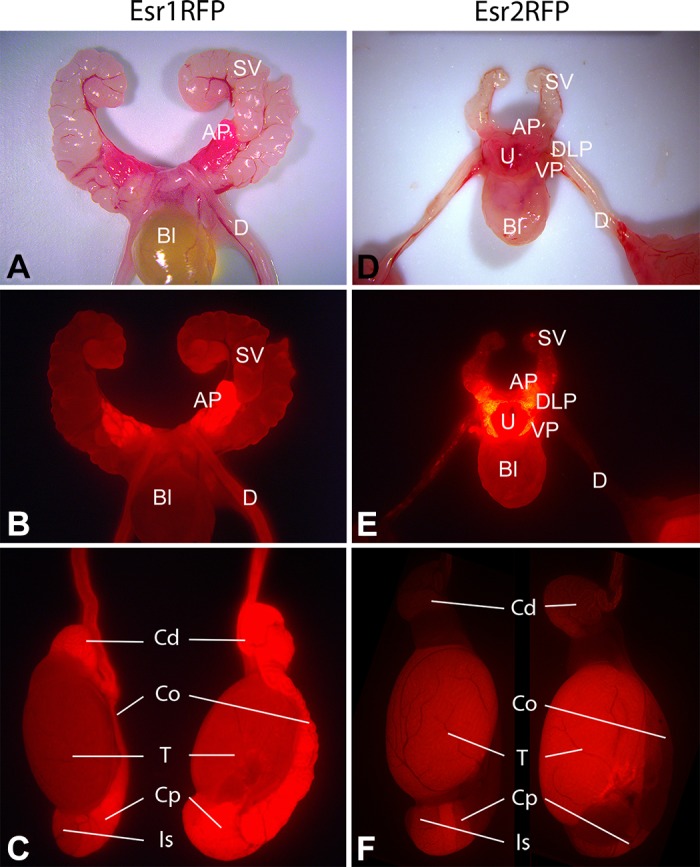
Estrogens have historically been associated with female reproduction, but work over the last two decades established that estrogens and their main nuclear receptors (ESR1 and ESR2) and G protein-coupled estrogen receptor (GPER) also regulate male reproductive and nonreproductive organs. 17β-Estradiol (E2) is measureable in blood of men and males of other species, but in rete testis fluids, E2 reaches concentrations normally found only in females and in some species nanomolar concentrations of estrone sulfate are found in semen. Aromatase, which converts androgens to estrogens, is expressed in Leydig cells, seminiferous epithelium, and other male organs. Early studies showed E2 binding in numerous male tissues, and ESR1 and ESR2 each show unique distributions and actions in males. Exogenous estrogen treatment produced male reproductive pathologies in laboratory animals and men, especially during development, and studies with transgenic mice with compromised estrogen signaling demonstrated an E2 role in normal male physiology. Efferent ductules and epididymal functions are dependent on estrogen signaling through ESR1, whose loss impaired ion transport and water reabsorption, resulting in abnormal sperm. Loss of ESR1 or aromatase also produces effects on nonreproductive targets such as brain, adipose, skeletal muscle, bone, cardiovascular, and immune tissues. Expression of GPER is extensive in male tracts, suggesting a possible role for E2 signaling through this receptor in male reproduction. Recent evidence also indicates that membrane ESR1 has critical roles in male reproduction. Thus estrogens are important physiological regulators in males, and future studies may reveal additional roles for estrogen signaling in various target tissues.


Figures

FIGURE 1.
FIGURE 1.
Aromatase (Cyp19) expression in male mouse reproductive tract. A: testis (T) and epididymis (E) from an adult (71-day-old) Cyp19RFP mouse showing RFP expression that is extensive within the testis, but lower in epididymis. B: adult testis showing immunohistochemical localization of aromatase in Leydig cells (L), round spermatids (Rs), and elongated spermatids (Es). C: adult caput epididymis showing immunohistochemical localization of aromatase in the cytoplasmic droplet (Cd) of sperm (Sp) in the tubular lumen. E, epithelium.
FIGURE 2.
FIGURE 2.
Expression of ESR1 and ESR2 in male mouse reproductive tract. Representative samples of immunohistochemical staining with 2 different ESR1 and ESR2 antibodies and red fluorescent protein (EsrRFP) mice. In Esr1RFP (63 days of age) and Esr2RFP mice (59 days of age), cell lineages expressing Esr1 or 2 will subsequently show RFP and may not correlate with current immunohistochemical staining. ESR1 staining is primarily nuclear using 6F11 [NCL-ER-6F11 antibody (Novocastra, Newcastle upon Tyne, UK)] and labels many more epididymal cell types than does the anti-ESR1 antibody 06–935 (Millipore, NH). Testis shows ESR1 exclusively in interstitial or Leydig cells (L) with no immunostaining in seminiferous tubules (St), although low fluorescence was seen with RFP. Efferent ductules show strong epithelial nuclear ESR1 staining with both antibodies, but cytoplasmic staining was also seen, especially with 06–935. In epididymis, apical (Ap) and clear cells (Cl) show strong nuclear staining with 6F11, but staining differences were observed in other cell types with the two antibodies. ESR2 staining was more widespread than ESR1. However, the S-40 ESR2 antibody (Dr. Saunders, Univ. of Edinburgh) showed intense nuclear staining, while PA1–311 (Thermo, Waltham, MA) shows considerable or exclusive cytoplasmic staining. The lack of RFP fluorescence in efferent ductules and most epididymal regions may indicate that ESR2 expression in these regions is delayed past day 60. In rats, ESR1 is expressed in efferent ducts and epididymis earlier than ESR2 (605), although in humans the opposite occurs (627). In the pig epididymis, ESR2 does not appear until after puberty (109). E, epithelium; Lu, lumen; Sm, smooth muscle; Ci, cilia. [Images for ESR1 using the 6F11 antibody and for ESR2 using the S-40 antibody were modified from Zhou et al. (763).]
FIGURE 3.
FIGURE 3.
Efferent duct expression of ESR1 in 3 mammalian species. Efferent ductules from mouse (A), marmoset monkey (B), and hamster (C) show intense immunostaining for ESR1. In most species, both ciliated (Ci) and nonciliated (Nc) cells have strong reactions in the nucleus, with some light cytoplasmic staining. However, the monkey ciliated cells were inconsistent, with some staining slightly positive and others being negative. The hamster image shows the efferent duct/initial segment junction, with intense staining of efferent duct epithelium but minimal epididymal staining.
FIGURE 4.
FIGURE 4.
Expression of ESR1 and ESR2 in male mouse reproductive tract visualized using EsrRFP mice. In Esr1RFP (A--C) and Esr2RFP (D--F) mice, red fluorescent protein (RFP) expression is under the control of the respective steroid receptor. A and B: whole mounts of adult (63-day-old) Esr1RFP reproductive tract photographed with normal light (A) or by exposing the tissue to light at 549 nm and then looking at fluorescence emission at 574 nm using a Zeiss HBO 100 illuminating system (B). The anterior prostate (AP; also called the coagulating gland) showed strong ESR1 expression (B) compared with much weaker ESR1 expression in seminal vesicles (SV) and ductus deferens (D), while expression in the bladder (Bl) was at the limit of detection. C: in whole mounts of adult (63-day-old) Esr1RFP testis and associated structures, the testis (T) is lightly positive, but more intense fluorescence is seen in the initial segment (Is), caput (Cp), corpus (Co), and cauda (Cd) regions of the epididymis. In juvenile (22-day-old) Esr2RFP male mice (D and E), ESR2 showed intense expression in ventral and dorsolateral prostate (VP and DLP, respectively), while AP and urethra (U) showed modest expression, Bl and SV showed minimal expression, and the ductus deferens (D) was essentially negative. F: in adult (59-day-old) Esr2RFP male mice, the IS and Cp of the epididymis showed clear signal for ESR2, while ESR2 expression in the Co and Cd regions of the epididymis were basically undetectable. In contrast to the epididymis, where ESR1-RFP expression (C) was more dominant compared with ESR2-RFP (F), in the testis ESR2-RFP expression (F) was stronger than ESR1-RFP expression (C).
FIGURE 5.
FIGURE 5.
Efferent ductule morphology in Esr1KO and anti-estrogen (Faslodex)-treated mice. A and B: light microscopy of adult wild-type (WT) and Esr1KO efferent ductules. WT ducts have a periodic acid (PAS)-positive brush border of microvilli (Mv) on nonciliated cells, which move sodium ions (Na+) and water (H2O) to concentrate luminal sperm that are transported into the epididymis. Long cilia (Ci) project into the lumen. Esr1KO ducts have a dilated lumen and reduced epithelial height. Epithelium is deficient in microvilli, and cilia are fewer and shorter. Sodium transport and water resorption are inhibited, but chloride ion (Cl-) secretion into the lumen is increased, adding to water accumulation. C--F: transmission electron microscopy of wild-type and Esr1KO efferent ductules. WT epithelium is taller than Esr1KO (double-headed red arrows). WT nonciliated cells (Nc) show a well-developed luminal border of microvilli (double-headed black arrows), coated pits (Cp), and apical resorption tubules (At). Esr1KO duct epithelium is short, and microvilli of nonciliated cells are short or absent and coated pits and apical tubules are reduced in apical cytoplasm. G--J: light microscopy of adult control and anti-estrogen (Faslodex)-treated efferent ductules. Control ducts have a smaller lumen but taller epithelium than Faslodex-treated mice. Sodium and water transport are actively moved into the interstitium but inhibited in treated epithelium. Nonciliated cells in controls have a PAS+ brush border of microvilli and ciliated cells support long cilia projecting into the lumen (I), in contrast to Faslodex-treated epithelia (J). K and L: transmission electron microscopy of control Faslodex-treated efferent ductules. Control epithelium is tall (K) compared with Faslodex-treated ducts (L). Control nonciliated cells have a well-developed luminal microvillous border, while treated duct epithelium has short microvilli. Control ciliated cells have numerous basal bodies (red arrowheads) in the apical cytoplasm to support cilia projecting into the lumen, in contrast to reduced cilia in treated cells (L). [The Esr1KO and Faslodex-treated images from Hess et al. (298), with permission from Taylor & Francis Group, LLC; and from Hess (289), with permission from the Brazilian College of Animal Reproduction.]
FIGURE 6.
FIGURE 6.
Estrogen synthesis and its targets in male reproductive tract. This figure summarizes the variation reported for the localization of estrogen receptors (ESR) in epithelia and stroma of testis, rete testis, efferent ductules, initial segment (seg), caput, cauda epididymis (epi), vas deferens, prostate, and other organs. Only nuclear ESR1 (yellow color) are represented. However, in some tissues, cytoplasmic and membrane ESRs have been documented. Receptor localization varies widely between species and with various antibodies (305). In adult testis, CYP19A1 (red color), the cytochrome P450 aromatase enzyme responsible for converting T to E2, is principally found in spermatids and mature sperm in seminiferous tubules and Leydig cells. These two sources of estrogen in the male reproductive system are directed to separate physiological pathways: 1) E2 from Leydig cells may target the seminiferous epithelium, although Sertoli and germ cells appear to be inconsistent in their ESR1 expression. This minor source of estrogen enters the blood and targets stromal and epithelial tissues not only in the reproductive tract but also all other ER-expression organs. 2) Germ cell production of E2 begins within seminiferous epithelium and continues with the localization of aromatase in the cytoplasmic droplet of spermatozoa transported in the lumen of the reproductive tract. The major target of luminal E2 is efferent ductule epithelium, where ESR1 expression is the highest in the body. The major function of efferent ductules is reabsorption of nearly 90% of the luminal fluid, which increases sperm concentrations entering the initial segment. This major physiological function, under ESR1 regulation, involves kidney-like physiology of the nonciliated cells (outlined in the red box), of which several genes are directly Esr1 regulated (296, 342).
FIGURE 7.
FIGURE 7.
Structural abnormalities of NOER mouse sperm arise in the post-seminiferous tubular environment. Forty-day-old NOER male testes were fixed and stained with Masson’s trichrome. A: seminiferous tubular epithelium at spermiation (stage VIII) shows normal sperm with straight tails. B: rete testis region of NOER mice shows high numbers of abnormal sperm with coiled tails (CT). SE, seminiferous epithelium; Es, elongated spermatids.
FIGURE 8.
FIGURE 8.
Efferent ductule epithelium from adult wild-type (WT), Esr1KO, and NOER (nuclear-only ESR1) mice. Periodic acid-Schiff (pink) and hematoxylin (blue) staining. Bar = 20 μm. A: WT epithelium is short columnar with ciliated (Ci) and nonciliated (Nc) cell. Nonciliated cells have a prominent brush border of microvilli (Mv) lining the lumen that contains diluted population of sperm (Sp). B: Esr1KO epithelium is shorter in height than WT, with significant loss of apical cytoplasm and much of the nonciliated microvillus border. Cilia numbers are reduced. C: NOER epithelium is shorter in height than WT, lacks a microvillus border, and shows reduced apical cytoplasm, similar to Esr1KO mice. Cilia also are reduced. Abnormal sperm with coiled tails are seen in the lumen.
FIGURE 9.
FIGURE 9.
Estrogen signaling pathways within prostatic epithelial cells. E2 and other agonists have multiple receptors and pathways that can be engaged to produce a variety of effects within cells. Both ESR1 and 2 (represented as ER) signal through classic genomic pathways. In addition, both ESR1 and 2 are present at the membrane and activate rapid signaling pathways upon ligand binding, including phosphorylation of Akt and/or the MAPK cascade. Multiple downstream effectors can be activated in a context-specific and perhaps ER-selective manner resulting in histone modifications (H3K4, H3K9, H3K27 trimethylation or demethylation) and direct transcriptional activation through intermediaries that include c-fos, c-jun, SP1, and NFkB as well as phosphorylation of nuclear ERs that amplify their activities. Finally, estrogens can signal through GPER, which activates PKA signaling.

References

    1. Acconcia F, Ascenzi P, Fabozzi G, Visca P, Marino M. S-palmitoylation modulates human estrogen receptor-alpha functions. Biochem Biophys Res Commun 316: 878–883, 2004. doi: 10.1016/j.bbrc.2004.02.129. - DOI - PubMed
    1. Adams JY, Leav I, Lau KM, Ho SM, Pflueger SM. Expression of estrogen receptor beta in the fetal, neonatal, and prepubertal human prostate. Prostate 52: 69–81, 2002. doi: 10.1002/pros.10103. - DOI - PubMed
    1. Adlanmerini M, Solinhac R, Abot A, Fabre A, Raymond-Letron I, Guihot AL, Boudou F, Sautier L, Vessières E, Kim SH, Lière P, Fontaine C, Krust A, Chambon P, Katzenellenbogen JA, Gourdy P, Shaul PW, Henrion D, Arnal JF, Lenfant F. Mutation of the palmitoylation site of estrogen receptor α in vivo reveals tissue-specific roles for membrane versus nuclear actions. Proc Natl Acad Sci USA 111: E283–E290, 2014. doi: 10.1073/pnas.1322057111. - DOI - PMC - PubMed
    1. Agarwal VR, Takayama K, Van Wyk JJ, Sasano H, Simpson ER, Bulun SE. Molecular basis of severe gynecomastia associated with aromatase expression in a fibrolamellar hepatocellular carcinoma. J Clin Endocrinol Metab 83: 1797–1800, 1998. - PubMed
    1. Aguirre JI, Plotkin LI, Gortazar AR, Millan MM, O’Brien CA, Manolagas SC, Bellido T. A novel ligand-independent function of the estrogen receptor is essential for osteocyte and osteoblast mechanotransduction. J Biol Chem 282: 25501–25508, 2007. doi: 10.1074/jbc.M702231200. - DOI - PubMed

Publication types

MeSH terms

LinkOut - more resources