. 2017 May 24;9(391):eaal4682.
doi: 10.1126/scitranslmed.aal4682.

The brain microenvironment mediates resistance in luminal breast cancer to PI3K inhibition through HER3 activation

Affiliations

The brain microenvironment mediates resistance in luminal breast cancer to PI3K inhibition through HER3 activation

David P Kodack et al. Sci Transl Med. .

Abstract

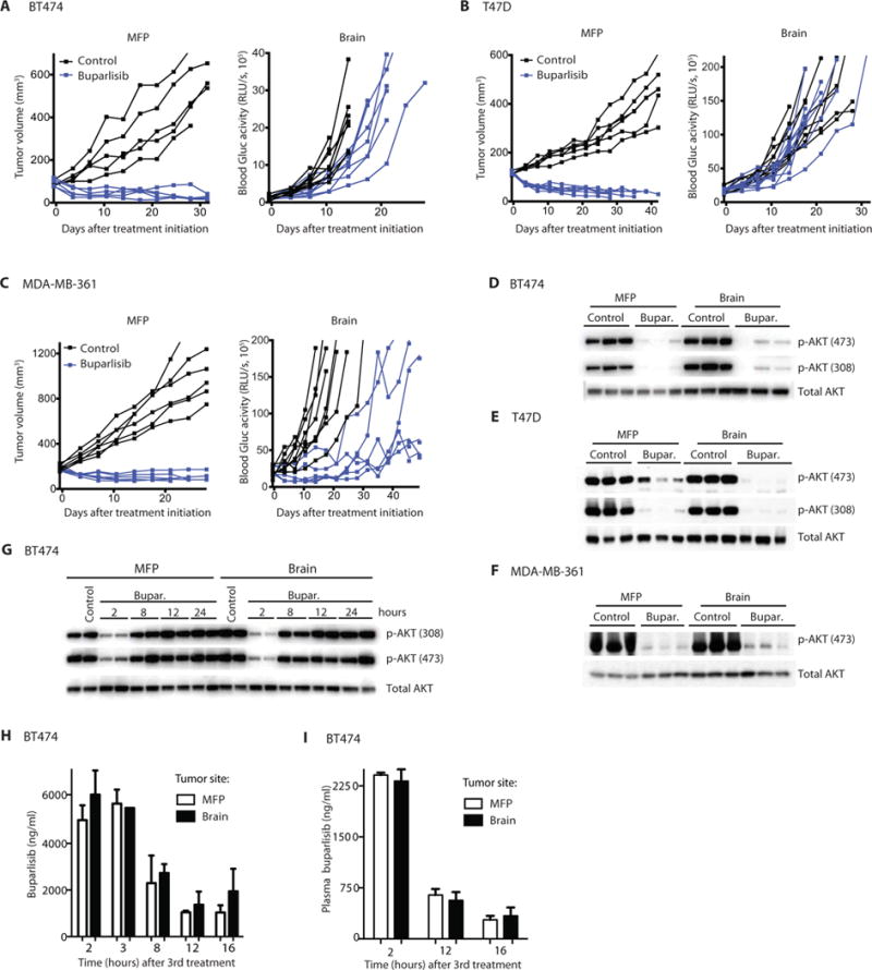
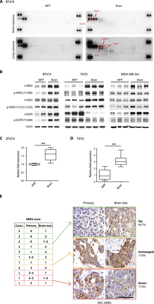
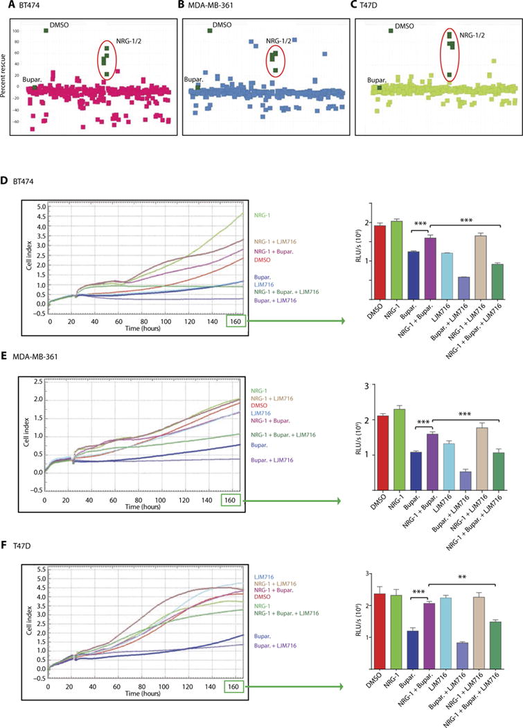
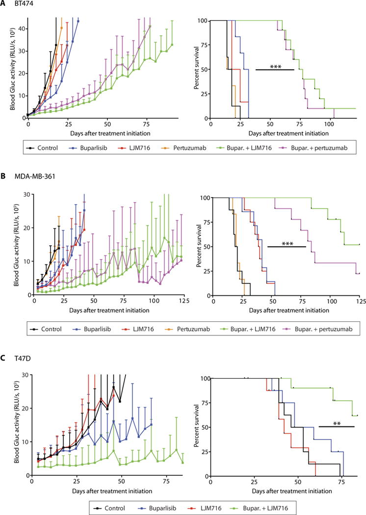
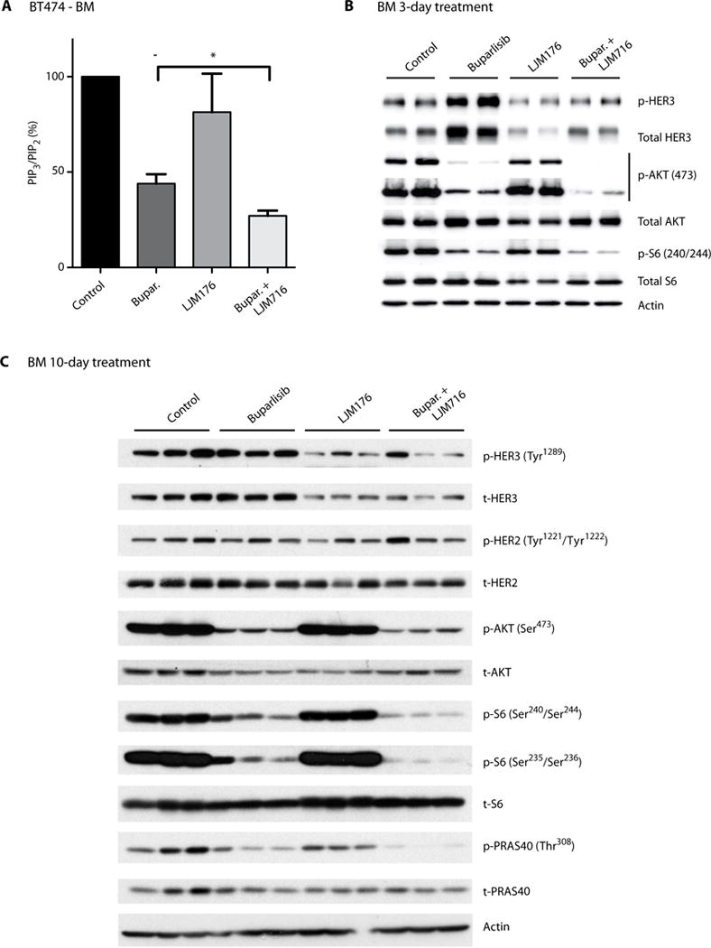
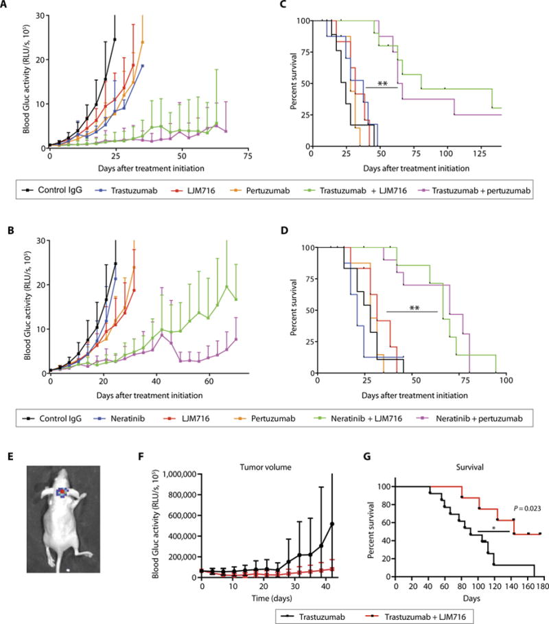
Although targeted therapies are often effective systemically, they fail to adequately control brain metastases. In preclinical models of breast cancer that faithfully recapitulate the disparate clinical responses in these microenvironments, we observed that brain metastases evade phosphatidylinositide 3-kinase (PI3K) inhibition despite drug accumulation in the brain lesions. In comparison to extracranial disease, we observed increased HER3 expression and phosphorylation in brain lesions. HER3 blockade overcame the resistance of HER2-amplified and/or PIK3CA-mutant breast cancer brain metastases to PI3K inhibitors, resulting in marked tumor growth delay and improvement in mouse survival. These data provide a mechanistic basis for therapeutic resistance in the brain microenvironment and identify translatable treatment strategies for HER2-amplified and/or PIK3CA-mutant breast cancer brain metastases.


Conflict of interest statement

Competing interests: R.K.J. received consultant fees from Enlight, Ophthotech, SPARC, and SynDevRx. R.K.J. owns equity in Enlight, Ophthotech, SynDevRx, and XTuit and serves on the Board of Directors of XTuit and the Boards of Trustees of Tekla Healthcare Investors, Tekla Life Sciences Investors, the Tekla Healthcare Opportunities Fund, and the Tekla World Healthcare Fund. J.A.E. is a paid consultant for Novartis, Sanofi-Aventis, AstraZeneca, Chugai, Amgen, Genentech, GSK, Merck, and Pfizer and holds equity in Gatekeeper Pharmaceuticals. J.A.E. also received research support from Novartis and is now an employee of Novartis. D.P.K., V.A., G.B.F., Q.S., D.F., and R.K.J. are inventors on patent application (PAT056561) held/submitted by D.P.K., V.A., G.B.F., Q.S., D.F., and R.K.J. (MGH/Harvard Medical School and Novartis) that covers the finding that a combination of a PI3K inhibitor and a Her3 antagonist can be used to treat BC BMs.

Figures

Fig. 1
Fig. 1. HER2-amplified and/or PIK3CA-mutant breast tumors display differential response to PI3K inhibition when grown in the MFP versus brain despite similar drug delivery
Established HER2-amplified BT474-Gluc (A), PIK3CA-mutant T47D-Gluc (B), and HER2-amplified and PIK3CA-mutant MDA-MB-361-Gluc tumors (C) growing in the MFP (left) or brain (right) were treated with buparlisib (BKM120) or vehicle (PEG 300), and tumor volume (MFP) or blood Gluc activity (brain) was measured at the indicated times (BT474: MFP n = 5, brain n = 7 to 9; MDA-MB-361: MFP n = 6, brain n = 6 to 7; T47D: MFP n = 6 to 7, brain n = 9 to 10). RLU/s, relative light units per second. BT474-Gluc (D), T47D-Gluc (E), or MDA-MB-361-Gluc (F) tumor tissue, collected 2 hours after the final treatment with buparlisib (Bupar.), was analyzed for AKT phosphorylation as a readout of PI3K inhibition. (G) BT474-Gluc tumor tissue, collected at the indicated time points after the third dose of buparlisib (48 hours after the first dose), was analyzed for AKT phosphorylation. (H) The concentration of buparlisib in BT474-Gluc tumor tissue collected after the indicated time points was determined [unpaired t test, 2 hours (n ≥ 4), P = 0.41; 8 hours (n = 2), P = 0.78; 12 hours (n = 2), P = 0.67; 16 hours (n ≥ 3), P = 0.45]. (I) Plasma concentration of buparlisib in mice whose tumor buparlisib concentration was analyzed in (H) [unpaired t test, 2 hours (n ≥ 4), P = 0.64; 12 hours (n = 2), P = 0.66; 16 hours (n = 4), P = 0.68]. Data are means ± SD.
Fig. 2
Fig. 2. HER3 is overexpressed and hyperactivated in BMs
(A) A phosphoreceptor tyrosine kinase array depicts ErbB family members as hyperphosphorylated in HER2-amplified BT474 breast tumors growing in the brain (right) compared with the MFP (left). (B) Breast tumor tissue from BT474, T47D, or MDA-MB-361 MFP or brain tumors was analyzed for phosphorylated and total (t) EGFR, HER2, and HER3. (C and D) Quantification of HER3 mRNA (human) in BT474 (n = 4) and T47D (brain; n = 7 and 9) (unpaired t test, **P < 0.001). Data are means ± SD. (E) Whole-tissue section immunohistochemistry (IHC) analysis of HER3 protein in matched human primary and brain metastatic (met.) HER2-positive breast tumor tissue (left). Representative IHC images of matched human primary and brain metastatic HER2-positive breast tumor tissue stained for HER3 (right). Percentage of HER3 signal in BM and matched primary tumor is depicted (scale bar, 200 μm).
Fig. 3
Fig. 3. NRGs induce in vitro resistance to PI3K inhibition in HER2-amplified and/or PIK3CA-mutant BC cells, and this is dependent on HER3 activation
(A to C) A library of secreted proteins was screened for their ability to rescue the inhibition of BT474 (A), MDA-MB-361 (B), and T47D (C) BC cell growth in vitro by buparlisib. Each dot represents the average of three replicates of cells conditioned with the same supernatant. “DMSO” represents cells treated with dimethyl sulfoxide control in the absence of secreted proteins (rescue, buparlisib-treated cells in the presence of growth factor relative to control). (D to F) In vitro xCELLigence growth assay of BT474 (D), MDA-MB-361 (E), or T47D (F) BC cells exposed under the indicated conditions for the indicated time. The y axis is cell index defined as the relative change in measured electrical impedance. The graphs on the right show CellTiter-Glo luminescence viability measurements at the end of the experiments [one-way analysis of variance (ANOVA)/Tukey test, **P = 0.0009, ***P < 0.0001]. Data are means ± SD.
Fig. 4
Fig. 4. Therapeutic targeting of HER3 sensitizes HER2-amplified and PI3KCA-mutant BC BMs to PI3K inhibition
Tumor growth curve (left) and survival analysis (right) of established BT474-Gluc (A), MDA-MB-361 (B), or T47D (C) brain tumors untreated or treated with buparlisib, LJM716, pertuzumab, buparlisib and LJM716, or buparlisib and pertuzumab [log-rank test; (A) BT474-Gluc—n = 6 to 10 per group; buparlisib + LJM716 versus buparlisib: hazard ratio (HR), 0.19; 95% confidence interval (CI), 0.04 to 0.94; P < 0.0001; buparlisib + pertuzumab versus buparlisib: HR, 0.20; 95% CI, 0.04 to 0.97; P < 0.0001; (B) MDA-MB-361—n = 6 to 9 per group; buparlisib + LJM716 versus buparlisib: HR, 0.13; 95% CI, 0.03 to 0.60; P < 0.0001; buparlisib + pertuzumab versus buparlisib: HR, 0.19; 95% CI, 0.04 to 0.78; P < 0.0001; (C) T47D—n = 8 to 11 per group; buparlisib + LJM716 versus buparlisib: HR, 0.19; 95% CI, 0.05 to 0.73; P = 0.005] (**P = 0.005 and ***P < 0.0001). Tumor size data are means ± SD.
Fig. 5
Fig. 5. HER3 blockade inhibits PI3K signaling in BM
(A) Analysis of PIP3/PIP2 ratio in HER2-amplified BT474-Gluc BM untreated or treated with buparlisib, LJM716, or buparlisib and LJM716. Tumor tissue was collected 3 hours after the 10th treatment with buparlisib (24-hour intervals) and/or fifth dose of LJM716 (48-hour intervals) (unpaired t test, n = 5, *P = 0.0178). Data are means ± SEM. (B) Analysis of HER3, AKT, and S6 phosphorylation in HER2-amplified BT474-Gluc BM untreated or treated for 3 days with buparlisib, LJM716, or their combination. (C) Analysis of HER3, HER2, AKT, S6, and PRAS phosphorylation in HER2-amplified BT474-Gluc brain tumors untreated or treated for 10 days with buparlisib, LJM716, or their combination.
Fig. 6
Fig. 6. Therapeutic targeting of HER3 sensitizes HER2-amplified BC BMs to HER2 inhibition
Tumor growth curve (A and B) and survival analysis (C and D) of established BT474-Gluc brain tumors untreated or treated with trastuzumab, neratinib, pertuzumab, LJM716, and indicated combinations (n = 8 to 10 per group; log-rank test; trastuzumab + LJM716 versus trastuzumab: HR, 0.16, 95% CI, 0.03 to 0.83; **P < 0.001; trastuzumab + pertuzumab versus trastuzumab: HR, 0.17; 95% CI, 0.04 to 0.85; **P < 0.001; neratinib + LJM716 versus neratinib: HR, 0.24; 95% CI, 0.06 to 0.90; P < 0.001; neratinib + pertuzumab versus neratinib: HR, 0.24; 95% CI, 0.06 to 0.91; P = 0.001). Tumor size data are means ± SD. IgG, immunoglobulin G. (E) Representative bioluminescence whole-body imaging of brain BT474-Gluc tumors from an intracarotid model. Tumor growth curve (F) and survival analysis (G) of BT474-Gluc brain tumors treated with trastuzumab (n = 13) or trastuzumab + LJM716 (n = 8) in the intracarotid model (log-rank test: HR, 0.30; 95% CI, 0.11 to 0.83; *P = 0.02).

References

    1. Kodack DP, Askoxylakis V, Ferraro GB, Fukumura D, Jain RK. Emerging strategies for treating brain metastases from breast cancer. Cancer Cell. 2015;27:163–175. - PMC - PubMed
    1. Olson EM, Abdel-Rasoul M, Maly J, Wu CS, Lin NU, Shapiro CL. Incidence and risk of central nervous system metastases as site of first recurrence in patients with HER2-positive breast cancer treated with adjuvant trastuzumab. Ann Oncol. 2013;24:1526–1533. - PMC - PubMed
    1. Murrell DH, Foster PJ, Chambers AF. Brain metastases from breast cancer: Lessons from experimental magnetic resonance imaging studies and clinical implications. J Mol Med. 2014;92:5–12. - PubMed
    1. Dasararaju R, Mehta A. Current advances in understanding and managing secondary brain metastasis. CNS Oncol. 2013;2:75–85. - PMC - PubMed
    1. Tamura K, Kurihara H, Yonemori K, Tsuda H, Suzuki J, Kono Y, Honda N, Kodaira M, Yamamoto H, Yunokawa M, Shimizu C, Hasegawa K, Kanayama Y, Nozaki S, Kinoshita T, Wada Y, Tazawa S, Takahashi K, Watanabe Y, Fujiwara Y. 64Cu-DOTA-trastuzumab PET imaging in patients with HER2-positive breast cancer. J Nucl Med. 2013;54:1869–1875. - PubMed

MeSH terms

Substances