. 2017 May 24;7(1):2320.
doi: 10.1038/s41598-017-02456-y.

An episomal vector-based CRISPR/Cas9 system for highly efficient gene knockout in human pluripotent stem cells

Affiliations

An episomal vector-based CRISPR/Cas9 system for highly efficient gene knockout in human pluripotent stem cells

Yifang Xie et al. Sci Rep. .

Erratum in

Abstract

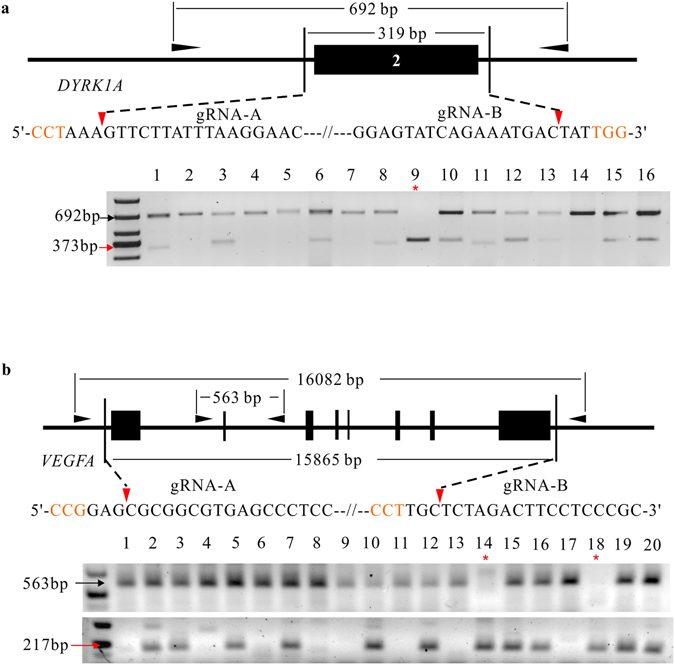
Human pluripotent stem cells (hPSCs) represent a unique opportunity for understanding the molecular mechanisms underlying complex traits and diseases. CRISPR/Cas9 is a powerful tool to introduce genetic mutations into the hPSCs for loss-of-function studies. Here, we developed an episomal vector-based CRISPR/Cas9 system, which we called epiCRISPR, for highly efficient gene knockout in hPSCs. The epiCRISPR system enables generation of up to 100% Insertion/Deletion (indel) rates. In addition, the epiCRISPR system enables efficient double-gene knockout and genomic deletion. To minimize off-target cleavage, we combined the episomal vector technology with double-nicking strategy and recent developed high fidelity Cas9. Thus the epiCRISPR system offers a highly efficient platform for genetic analysis in hPSCs.


Conflict of interest statement

The authors declare that they have no competing interests.

Figures

Figure 1
Figure 1
An epiCRISPR system for exogenous gene-free genome editing in hPSCs. (a) Schematic of the epiCRISPR system design. The vector contains a U6 promoter-driven gRNA scaffold, an EF1a promoter-driven Cas9 fused to puromycin resistance gene and GFP with P2A peptides, and OriP/EBNA1 elements for the plasmid replication in eukaryocytes. Puro, Puromycin resistance gene. (b) Schematic of genome editing with epiCRISPR system. The epiCRISPR vector is transfected into hPSCs followed by drug selection. Only the transfected cells can survive and proliferate. The epiCRISPR vector can replicate in hPSCs and can be partitioned to daughter cells. In the absence of drug selection, the epiCRISPR vector can be lost, allowing edited cells free of exogenous gene expression. (c) The epiCRISPR system for stable gene expression in hPSCs. The left panel shows the epiCRISPR vector delivered into hPSCs with lipid-mediated transfection. The middle panel shows the epiCRISPR vector can replicate during hPSC proliferation under drug selection. The right panel shows the single cell-derived clones lost epiCRISPR vector without drug selection. (d) Episomal vector was decreased within cells over time after withdrawing puromycin selection. The plasmid was tested every two days by quantitative PCR (n = 3, error bars show mean ± S.D.). (e) The absence of episomal vector in single cell-derived hESC clones was confirmed by PCR targeting ampicillin sequence. Ctr1 is the positive control amplified from the epiCRISPR plasmid DNA; Ctr2 is the positive control amplified from the pooled hESCs containing epiCRISPR vector.
Figure 2
Figure 2
The epiCRISPR system for efficient gene knockout. (a) Schematic representation of the experimental procedure. (b) RFLP analysis of the indel rates generated by the epiCRISPR system at DYRK1A, EMX1, AAVS1, VEGFA, APC1 and MLH1 loci in hPSCs (n = 3, error bars show mean ± S.D.). (c) Representative indel sequences at EMX1 and MLH1 sites in hESCs. The AgeI and XbaI restriction sequences are shown in red. The red triangles indicate Cas9 cutting site. (d) RFLP analysis of single hESC-derived clones modified at AAVS1 locus. Ctr1 is the PCR band from unmodified cells without digestion. Ctr2 is the PCR band from unmodified cells with SacI digestion. The red triangle indicates the epiCRISPR-modified PCR bands; the black triangle indicates unmodified PCR bands.
Figure 3
Figure 3
The epiCRISPR system for efficient double-gene knockout. (a) Expression of two gRNAs on the epiCRISPR vector for double-gene knockout. (b) RFLP analysis of the indel rates generated by the epiCRISPR system with gRNA multiplexed targeting DYRK1A & EMX1, AAVS1 & VEGFA and APC1 & MLH1 in hPSCs (n = 3, error bars show mean ± S.D.). (c) RFLP analysis of single cell-derived clones multiplexed targeting DYRK1A and EMX1 loci. Ctr1 is the PCR band from unmodified cells without digestion. Ctr2 is the PCR band from unmodified cells with digestion (BstXI for DYRK1A and AgeI for EMX1). Red triangles indicate the epiCRISPR-modified PCR bands; black triangles indicate unmodified PCR bands; the red asterisks indicate the homozygous knockout for both genes.
Figure 4
Figure 4
The epiCRISPR system for efficient genomic deletions. (a) The strategy for deletion of the 2nd exon of DYRK1A. A 373-bp PCR band will be present when deletion occurs (indicate by red arrow). A representative gel picture was shown. (b) The strategy for deletion of the full-coding sequence of VEGFA. Two pairs of primers were designed. One pair is to detect the non-deletion allele (563-bp) and one pair is to detect deletion events (217-bp, indicate by red arrow). The black triangles indicate the primers for the genotyping PCR reaction. The outer primers work only when deletion occurs. The PAM sequence is shown in orange; the red triangles indicate Cas9 cutting site; red arrows indicate the epiCRISPR-modified PCR bands; black arrows indicate unmodified PCR bands; the red asterisks indicate the homozygous knockout for both genes.
Figure 5
Figure 5
Analysis of off-target cleavage by targeted deep-sequencing. (a) The potential off-target sequences are shown on the left; the indel rates are shown on the right. On, on target sequence; OT, off-target sequence. The unmatched nucleotides are shown in red. (b) RFLP analysis of eSpCas9 cleavage at AAVS1, EMX1 and MLH1 sites. Red triangles indicate the epiCRISPR-modified PCR bands; black triangles indicate the unmodified PCR bands.
Figure 6
Figure 6
An epiCRISPRn system for efficient double-nicking. (a) Schematic of the epiCRISPRn vector design. A D10A mutation was introduced into the epiCRIPSR vector. (b) RFLP analysis of the indel rates generated by the epiCRISPRn system targeting DYRK1A, EMX1, AAVS1, VEGFA, APC1 and MLH1 loci (n = 3, error bars show mean ± S.D.). (c) Representative indel sequences generated by the epiCRISPRn at APC1 and MLH1 loci. The size of indels is shown on the right. The gRNA sequence is underlined; the PAM sequence is shown in orange; the restriction sites are shown in red, the red triangles indicate Cas9 cutting site.

Similar articles

Cited by

References

    1. Thomson JA, et al. Embryonic stem cell lines derived from human blastocysts. Science. 1998;282:1145–1147. doi: 10.1126/science.282.5391.1145. - DOI - PubMed
    1. Takahashi K, et al. Induction of pluripotent stem cells from adult human fibroblasts by defined factors. Cell. 2007;131:861–872. doi: 10.1016/j.cell.2007.11.019. - DOI - PubMed
    1. Mali P, et al. RNA-guided human genome engineering via Cas9. Science. 2013;339:823–826. doi: 10.1126/science.1232033. - DOI - PMC - PubMed
    1. Cong L, et al. Multiplex genome engineering using CRISPR/Cas systems. Science. 2013;339:819–823. doi: 10.1126/science.1231143. - DOI - PMC - PubMed
    1. Doudna JA, Charpentier E. Genome editing. The new frontier of genome engineering with CRISPR-Cas9. Science. 2014;346:1258096–1258096. doi: 10.1126/science.1258096. - DOI - PubMed

Publication types