. 2017 May 24;7(1):2390.
doi: 10.1038/s41598-017-02549-8.

The role of retinol dehydrogenase 10 in the cone visual cycle

Affiliations

The role of retinol dehydrogenase 10 in the cone visual cycle

Yunlu Xue et al. Sci Rep. .

Abstract

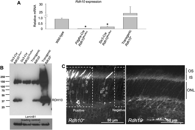
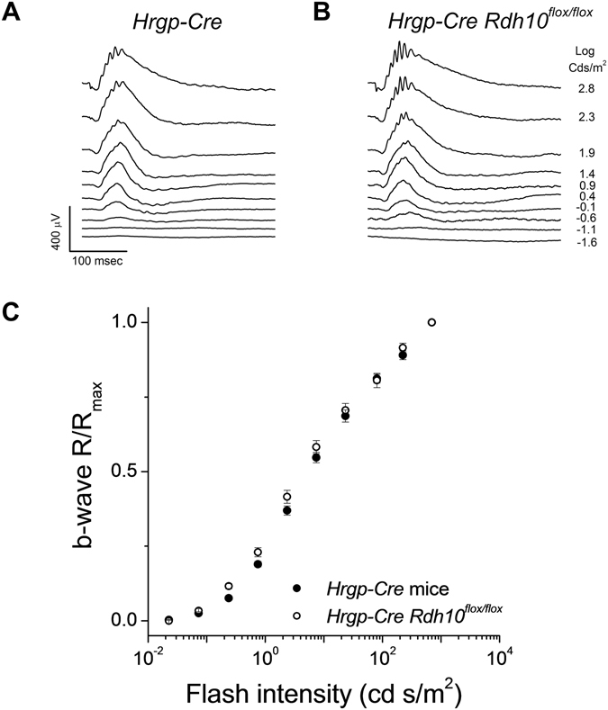
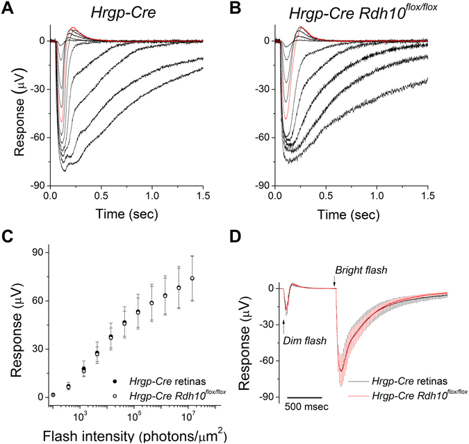
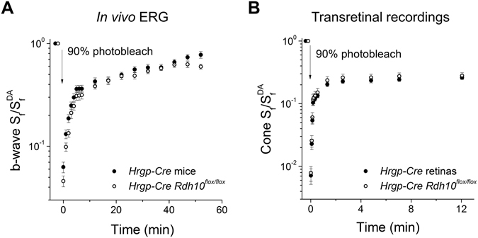
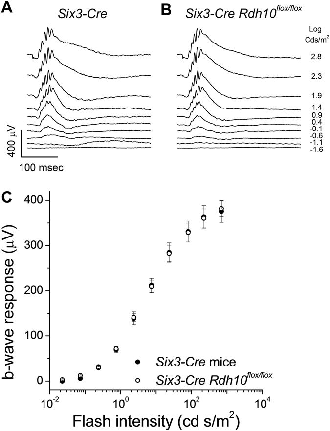
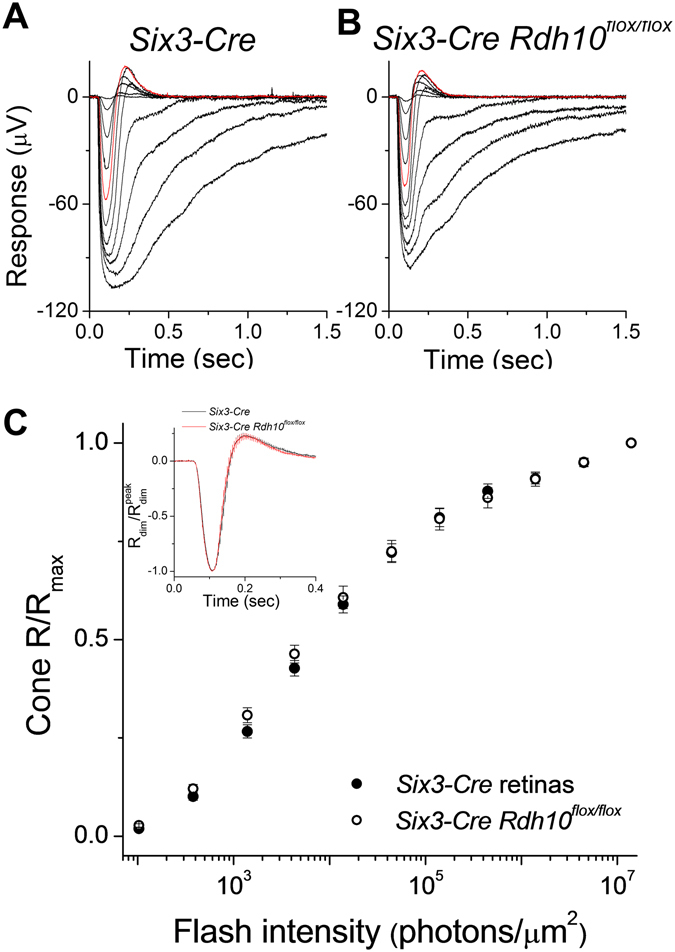
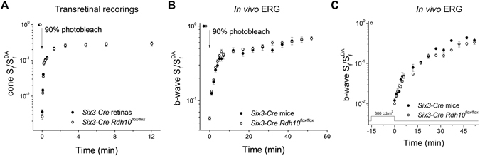
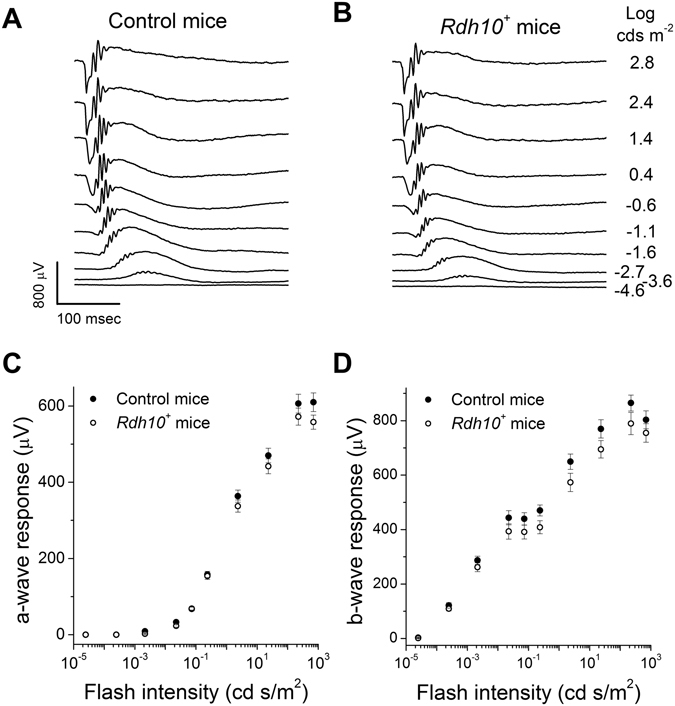
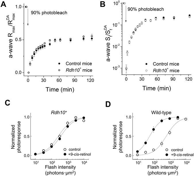
Pigment regeneration is critical for the function of cone photoreceptors in bright and rapidly-changing light conditions. This process is facilitated by the recently-characterized retina visual cycle, in which Müller cells recycle spent all-trans-retinol visual chromophore back to 11-cis-retinol. This 11-cis-retinol is oxidized selectively in cones to the 11-cis-retinal used for pigment regeneration. However, the enzyme responsible for the oxidation of 11-cis-retinol remains unknown. Here, we sought to determine whether retinol dehydrogenase 10 (RDH10), upregulated in rod/cone hybrid retinas and expressed abundantly in Müller cells, is the enzyme that drives this reaction. We created mice lacking RDH10 either in cone photoreceptors, Müller cells, or the entire retina. In vivo electroretinography and transretinal recordings revealed normal cone photoresponses in all RDH10-deficient mouse lines. Notably, their cone-driven dark adaptation both in vivo and in isolated retina was unaffected, indicating that RDH10 is not required for the function of the retina visual cycle. We also generated transgenic mice expressing RDH10 ectopically in rod cells. However, rod dark adaptation was unaffected by the expression of RDH10 and transgenic rods were unable to use cis-retinol for pigment regeneration. We conclude that RDH10 is not the dominant retina 11-cis-RDH, leaving its primary function in the retina unknown.


Conflict of interest statement

The authors declare that they have no competing interests.

Figures

Figure 1
Figure 1
RDH10 expression in different mouse lines. (A) mRNA analysis of Rdh10 expression in wild-type, Müller cell-specific RDH10 knockout (Pdgfra-Cre Rdh10 flox/flox), retina-specific RDH10 knockout (Six-Cre Rdh10 flox/flox), and RDH10 transgenic (Rdh10 +) mice. Data here and in all subsequent figures are shown as mean ± SEM. (B) Immunoblotting of Six3-Cre control, Six3-Cre Rdh10 flox/flox, transgenic Rdh10 (wild-type), and transgenic Rdh10 + retinas. (C) Retinal section from a transgenic Rdh10 + (left) and Rdh10 (wild-type) mice. The mosaic RDH10 expression in rods is shown in white and RDH10-positive and –negative sections are delineated by a box. OS, outer segment layer; IS, inner segment layer; ONL, outer nuclear layer.
Figure 2
Figure 2
Deletion of RDH10 in cones does not affect the photopic ERG b-wave responses in mice. Representative in vivo ERG responses from (A) Hrgp-Cre control, and (B) cone Rdh10 knockout (Hrgp-Cre Rdh10 flox/flox) mice. (C) Ensemble-averaged normalized cone b-wave responses of Hrgp-Cre control mice (n = 18, filled symbols) and Hrgp-Cre Rdh10 flox/flox mice (n = 18, open symbols) as a function of flash intensity. All mice were in Gnat1 −/− background to facilitate cone recordings.
Figure 3
Figure 3
Deletion of RDH10 in cone cells does not affect cone responses. Representative transretinal cone responses from (A) Hrgp-Cre control retinas, and (B) Hrgp-Cre Rdh10 flox/flox retinas. Red traces: responses to a 1.4 × 104 photons/μm2 test flash. (C) Ensemble-averaged intensity-response curves of Hrgp-Cre control cones (n = 12, filled symbols) and Hrgp-Cre Rdh10 flox/flox cones (n=12, open symbols). (D) Ensemble-averaged dim flash responses (flash intensity: 1.4 × 103 photons/μm) and bright flash responses (flash intensity: 4.5 × 106 photons/μm2) of Hrgp-Cre control cones (n = 12, black trace) and Hrgp-Cre Rdh10 flox/flox cones (n = 12, red trace). All mice were in Gnat1 −/− background to facilitate cone recordings.
Figure 4
Figure 4
Deletion of RDH10 in cones does not affect cone dark adaptation. (A) Normalized cone b-wave sensitivity (b-wave Sf/b-wave Sf DA) recovery following 90% pigment photobleach from HrgpP-Cre control (n = 18, filled symbols) and Hrgp-Cre Rdh10 flox/flox (n = 18, open symbols) mice measured by in vivo ERG recordings. (B) Normalized cone sensitivity (Sf/Sf DA) recovery following 90% pigment photobleach in Hrgp-Cre control (n = 13, filled symbols) and Hrgp-Cre Rdh10 flox/flox (n = 13, open symbols) isolated retinas measured by transretinal recordings. All mice were in Gnat1 −/− background to facilitate cone recordings.
Figure 5
Figure 5
Deletion of RDH10 in the entire retina does not affect the photopic ERG b-wave responses in mice. Representative in vivo ERG responses of (A) Six3-Cre control and (B) retina Rdh10 knockout (Six3-Cre Rdh10 flox/flox) mice. (C) Ensemble-averaged cone b-wave intensity-response curves of Six3-Cre control mice (n = 12, filled symbols) and Six3-Cre Rdh10 flox/flox mice (n = 10, open symbols). All mice were in Gnat1 −/− background to facilitate cone recordings.
Figure 6
Figure 6
Deletion of RDH10 in the entire retina does not affect cone responses. Representative transretinal cone responses from (A) Six3-Cre control, and (B) Six3-Cre Rdh10 flox/flox retinas. Red traces: responses to a 1.4 × 104 photons/μm2 test flash. (C) Normalized cone intensity-response curves of Six3-Cre control (n = 9, filled symbols) and Six3-Cre Rdh10 flox/flox (n = 8, open symbols) retinas. Inset: normalized cone dim flash responses from Six3-Cre control (n = 9, black trace) and Six3-Cre Rdh10 flox/flox (n = 8, red trace) retinas. All mice were in Gnat1 −/− background to facilitate cone recordings.
Figure 7
Figure 7
Deletion of RDH10 in the entire retina does not affect cone dark adaptation. (A) Normalized cone sensitivity (Sf/Sf DA) recovery following 90% pigment photobleach of Six3-Cre control (n = 9, filled symbols) and Six3-Cre Rdh10 flox/flox (n = 8, open symbols) retinas measured by transretinal recordings. (B) Normalized cone b-wave sensitivity (b-wave Sf/b-wave Sf DA) recovery following a 90% pigment photobleach of Six3-Cre control (n = 12, filled symbols) and Six3-Cre Rdh10 flox/flox (n = 10, open symbols) mice measured by in vivo ERG recordings. (C) Normalized cone b-wave sensitivity (b-wave Sf/b-wave Sf DA) recovery following a 15 min exposure to 300 cd/m2 Ganzfeld background light (530 nm wavelength) from Six3-Cre control (n = 4, filled symbols) and Six3-Cre Rdh10 flox/flox (n = 4, open symbols) mice measured by in vivo ERG recordings. All mice were in Gnat1 −/− background to facilitate cone recordings.
Figure 8
Figure 8
Ectopic expression of RDH10 in rod photoreceptor cells does not affect their photoresponses. Representative scotopic in vivo ERG responses from (A) control, and (B) transgenic Rdh10 (Rdh10 +) mice. (C) Ensemble-averaged scotopic a-wave responses (C) and scotopic b-wave responses (D) of control (n = 9, filled symbols) and Rdh10 + (n = 9, open symbols) mice measured by in vivo ERG recordings.
Figure 9
Figure 9
Ectopic expression of RDH10 does not enable rods to dark adapt faster or to oxidize cis-retinol for pigment regeneration. (A and B) Normalized rod scotopic a-wave maximal response (Rmax/Rmax DA; A) or sensitivity (Sf/Sf DA; B) recovery following a 90% pigment photobleach in control (n = 9, filled symbols) and Rdh10 + (n = 9, open symbols) mice measured by in vivo ERG recordings. (C and D) Normalized intensity-response curves determined by single-cell suction recordings from (C) transgenic Rdh10 + rods incubated in control solution (n = 13, open symbols) or in 100 µM 9-cis-retinol solution (n = 14, filled symbols), or from (D) wild-type rods incubated in control solution (n = 6, open symbols) or in 100 µM 9-cis-retinal solution (n = 11, filled symbols) following 50% pigment photobleach.

Similar articles

Cited by

References

    1. Ebrey T, Koutalos Y. Vertebrate Photoreceptors. Prog. Retin. Eye Res. 2001;20:49–94. doi: 10.1016/S1350-9462(00)00014-8. - DOI - PubMed
    1. Burns ME, Baylor DA. Activation, deactivation, and adaptation in vertebrate photoreceptor cells. Annu. Rev. Neurosci. 2001;24:779–805. doi: 10.1146/annurev.neuro.24.1.779. - DOI - PubMed
    1. Saari JC. Vitamin A metabolism in rod and cone visual cycles. Annu. Rev. Nutr. 2012;32:125–45. doi: 10.1146/annurev-nutr-071811-150748. - DOI - PubMed
    1. Lamb TD, Pugh EN. Dark adaptation and the retinoid cycle of vision. Prog. Retin. Eye Res. 2004;23:307–80. doi: 10.1016/j.preteyeres.2004.03.001. - DOI - PubMed
    1. Wang J, et al. Chromophore supply rate-limits mammalian photoreceptor dark adaptation. J. Neurosci. 2014;34:11212–21. doi: 10.1523/JNEUROSCI.1245-14.2014. - DOI - PMC - PubMed

Publication types

MeSH terms