Review
. 2011 Sep 26:3:29-47.
doi: 10.2147/EB.S16192. eCollection 2011.

Mitochondrial disorders and the eye

Affiliations
Review

Mitochondrial disorders and the eye

Nicole J Van Bergen et al. Eye Brain. .

Abstract

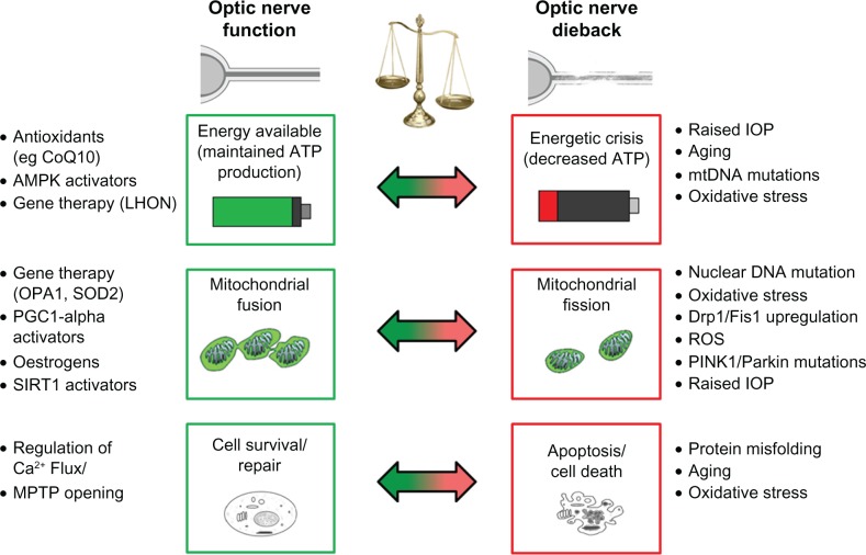
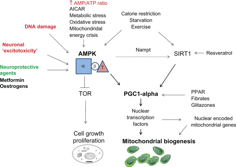
The clinical significance of disturbed mitochondrial function in the eye has emerged since mitochondrial DNA (mtDNA) mutation was described in Leber's hereditary optic neuropathy. The spectrum of mitochondrial dysfunction has become apparent through increased understanding of the contribution of nuclear and somatic mtDNA mutations to mitochondrial dynamics and function. Common ophthalmic manifestations of mitochondrial dysfunction include optic atrophy, pigmentary retinopathy, and ophthalmoplegia. The majority of patients with ocular manifestations of mitochondrial disease also have variable central and peripheral nervous system involvement. Mitochondrial dysfunction has recently been associated with age-related retinal disease including macular degeneration and glaucoma. Therefore, therapeutic targets directed at promoting mitochondrial biogenesis and function offer a potential to both preserve retinal function and attenuate neurodegenerative processes.

Keywords: aging; disease; eye; mitochondria; neuroprotection; retina.


Conflict of interest statement

Disclosure The authors report no conflicts of interest in this work.

Figures

Figure 1
Figure 1
Factors contributing to optic nerve degeneration. Note: The spectrum of mitochondrial dysfunction in the eye has emerged through understanding of the contribution of multiple stressors leading to mitochondrial failure and optic nerve loss. While controversy continues as to whether mitochondrial impairment in these diseases is primary or secondary to upstream disease pathways, the mitochondrion is emerging as a central culprit in cell death. Therapeutic developments aimed at boosting mitochondrial function are gathering pace. These approaches are likely to be of benefit not only in retinal disease, but also in diseases of the aging brain due to the growing evidence for a role of mitochondrial decline in age-related neurodegeneration.
Figure 2
Figure 2
Role of AMPK in neuroprotection. Note: AMP-Activated protein kinase (AMPK) is a heterotrimic enzyme that activates catabolic pathways and turns off anabolic pathways in response to increased AMP/ATP ratio (hypoxia,glucose deprivation, OXPHOS inhibition), Ca2+ mediated neuronal excitotoxicity, DNA damage. Low ATP levels activate AMPK by phosphorylation of AMPK at Thr172 in the kinase domain (in the alpha subunit). Once activated, AMPK inhibits ATP consumption, inhibits protein synthesis (by inhibiting Target of Rapamycin, TOR), and inhibits fatty acid and cholesterol synthesis. AMPK can drive mitochondrial biogenesis by phosphorylation of mitochondrial transcriptional coregulators including PGC-1a; and indirectly promote SIRT1 activity via regeneration of NAD+ through the enzyme Nampt. Emerging neuroprotective avenues for AMPK activation act via increasing AMP/ATP ratio (Glitazones), Metformin, and Estradiol.

References

    1. Wallace DC, Singh G, Lott MT, et al. Mitochondrial DNA mutation associated with Leber’s hereditary optic neuropathy. Science. 1988;242(4884):1427–1430. - PubMed
    1. Carelli V, La Morgia C, Valentino ML, Barboni P, Ross-Cisneros FN, Sadun AA. Retinal ganglion cell neurodegeneration in mitochondrial inherited disorders. Biochim Biophys Acta. 2009;1787(5):518–528. - PubMed
    1. Yu-Wai-Man P, Griffiths PG, Chinnery PF. Mitochondrial optic neuropathies – disease mechanisms and therapeutic strategies. Prog Retin Eye Res. 2011;30(2):81–114. - PMC - PubMed
    1. Fraser JA, Biousse V, Newman NJ. The neuro-ophthalmology of mitochondrial disease. Surv Ophthalmol. 2010;55(4):299–334. - PMC - PubMed
    1. Crouch PJ, Cimdins K, Duce JA, Bush AI, Trounce IA. Mitochondria in aging and Alzheimer’s disease. Rejuvenation Res. 2007;10(3):349–357. - PubMed