. 2016 May 20:8:55-65.
doi: 10.2147/EB.S94443. eCollection 2016.

Targeting VEGF in canine oxygen-induced retinopathy - a model for human retinopathy of prematurity

Affiliations

Targeting VEGF in canine oxygen-induced retinopathy - a model for human retinopathy of prematurity

D Scott McLeod et al. Eye Brain. .

Abstract

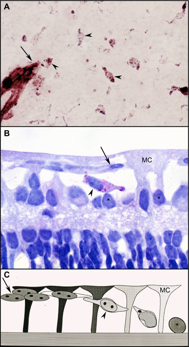
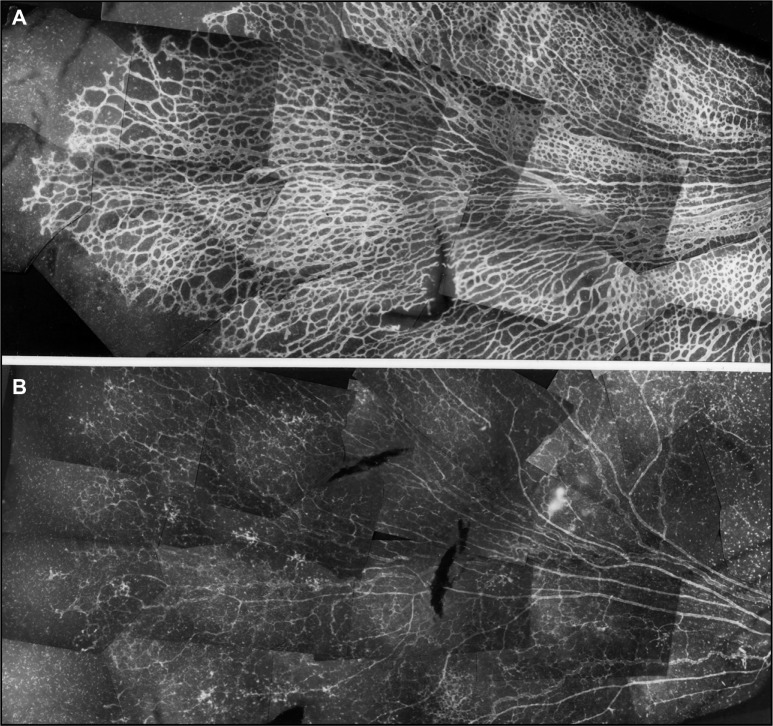
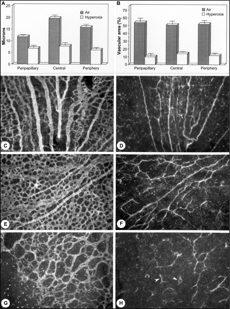
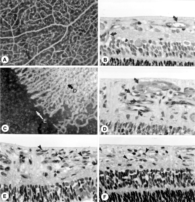
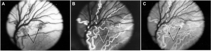
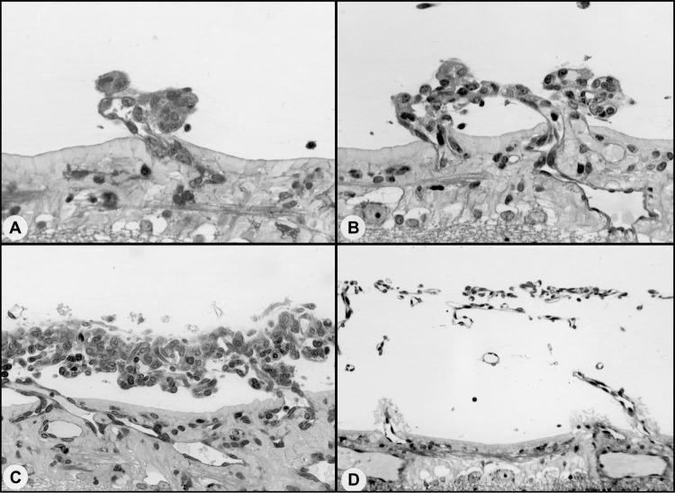
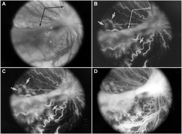
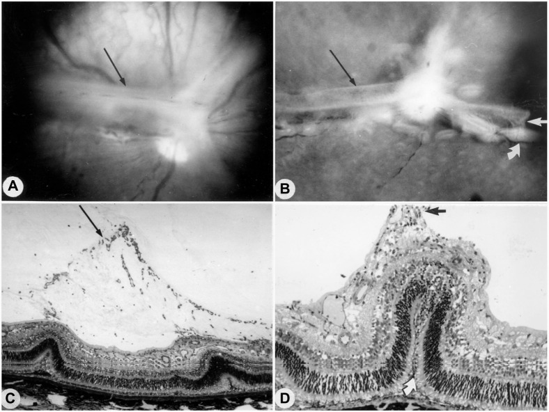
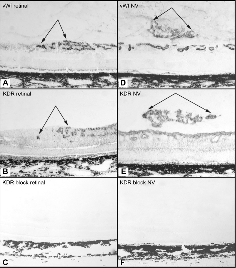
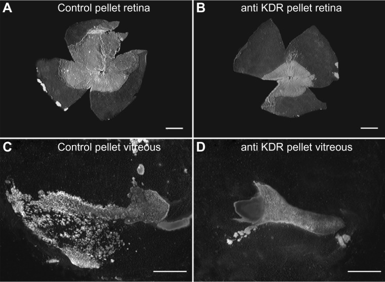
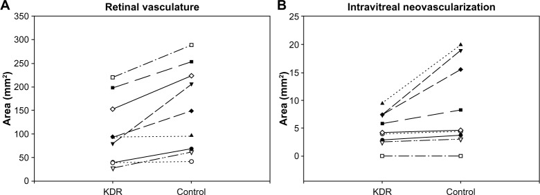
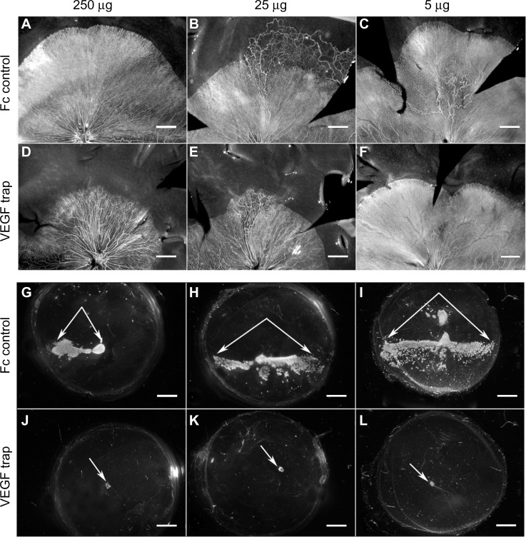
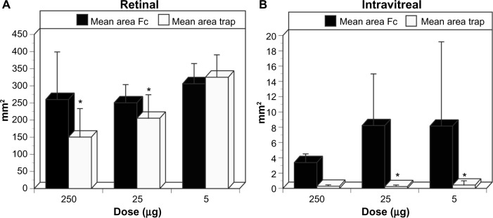
Development of the dog superficial retinal vasculature is similar to the mechanism of human retinal vasculature development; they both develop by vasculogenesis, differentiation, and assembly of vascular precursors called angioblasts. Canine oxygen-induced retinopathy (OIR) was first developed by Arnall Patz in an effort to experimentally determine the effects of hyperoxia on the development of the retinal vasculature. The canine OIR model has many characteristics in common with human retinopathy of prematurity. Exposure of 1-day-old dogs to hyperoxia for 4 days causes a vaso-obliteration throughout the retina. Vasoproliferation, after the animals have returned to room air, is robust. The initial small preretinal neovascular formations anastomose to form large preretinal membranes that eventually cause tractional retinal folds. The end-stage pathology of the canine model is similar to stage IV human retinopathy of prematurity. Therefore, canine OIR is an excellent forum to evaluate the response to drugs targeting VEGF and its receptors. Evaluation of an antibody to VEGF-R2 and the VEGF-Trap demonstrated that doses should be titered down so that preretinal neovascularization is inhibited but retinal revascularization is able to proceed, vascularizing peripheral retina and preventing it from being a source of VEGF.

Keywords: angioblasts; blood vessels; endothelial cells; oxygen; retina; retinopathy; vascular endothelial cell growth factor.


Conflict of interest statement

Disclosure The authors report no other conflict of interest in this work.

Figures

Figure 1
Figure 1
Vasculogenesis in the newborn dog. Notes: (A) ADPase flat mount showing vascular cords at the edge of developing vasculature (arrow) and angioblasts in a vascular periphery (arrowheads). (B) JB-4 section of ADPase flat-embedded 1-day-old dog retina at the edge of developing vasculature shows a vascular cord (arrow), Muller cell processes (MC), and a differentiating angioblast migrating within the cell-free spaces (arrowhead). Undifferentiated angioblasts are shown below the forming vascular cords (asterisks). (C) Schematic of angioblast differentiation in cell-free spaces made by MC: arrow, vascular cord; arrowhead, differentiating angioblast; asterisk, undifferentiated angioblast. Abbreviation: ADPase, adenosine diphosphatase.
Figure 2
Figure 2
Vaso-obliteration in the dog model of OIR. Notes: ADPase flat-embedded retinas of a 5-day-old air-control dog (A) and a 5-day-old dog after 4 days of hyperoxia (B). The majority of the capillaries have been obliterated. Magnification is ×14. Copyright © 1996 Association for Research in Vision and Ophthalmology. Reproduced from McLeod DS, Brownstein R, Lutty GA. Vaso-obliteration in the canine model of oxygen-induced retinopathy. Invest Ophthalmol Vis Sci. 1996;37:300–311. Abbreviations: OIR, oxygen-induced retinopathy; ADPase, adenosine diphosphatase.
Figure 3
Figure 3
Hyperoxia affects all regions uniformly in the dog OIR model. Notes: Lumenal diameters (A) and percent vascular area measurements (B) in the peripapillary, midperipheral, and peripheral regions of air controls (C, E, and G) versus oxygen-treated animals (D, F, and H). Arrowheads: surviving vascular endothelial cells and angioblasts. Magnification is ×60. Copyright © 1996 Association for Research in Vision and Ophthalmology. Reproduced from McLeod DS, Brownstein R, Lutty GA. Vaso-obliteration in the canine model of oxygen-induced retinopathy. Invest Ophthalmol Vis Sci. 1996;37:300–311. Abbreviation: OIR, oxygen-induced retinopathy.
Figure 4
Figure 4
Vasoproliferative phase of OIR in dog. Notes: Comparison of 22-day-old air control (A and B) and a 22-day-old oxygen-treated animals (CF) in flat perspective (A and C) and histologic sections (B, DF). In the air-control animals, inner retinal vasculature is complete and secondary capillaries are formed (curved arrows in B). In the oxygen-treated animals, a dense irregular pattern of broad capillaries has formed at the anterior border of revascularized retina (curved arrow in D), whereas a substantial area of retina remains avascular (straight arrow in C). Sections taken from the border in the oxygen-treated animal show multiple layers of capillaries at the vascular border (curved arrows in D), and numerous astrocytes in advance (arrowheads in E and F), which filled in the extracellular spaces present during normal vasculogenesis. (Magnifications: A=×100, B=×485, C=×100, D–F=×485). Copyright © 1996 Association for Research in Vision and Ophthalmology. Reproduced from McLeod DS, Crone SN, Lutty GA. Vasoproliferation in the neonatal dog model of oxygen-induced retinopathy. Invest Ophthalmol Vis Sci. 1996;37:1322–1333. Abbreviation: OIR, oxygen-induced retinopathy.
Figure 5
Figure 5
Intravitreal neovascularization in the dog OIR model. Notes: Red-free photograph (A) and early arterial-phase fluorescein angiogram (B and C) showing isolated intravitreal neovascular formations (paired arrows) in the posterior pole of a 45-day-old oxygen-treated animal. These formations are fed by a tortuous retinal artery. Copyright © 1998 Association for Research in Vision and Ophthalmology. Reproduced from McLeod DS, D’Anna SA, Lutty GA. Clinical and histopathologic features of canine oxygen-induced proliferative retinopathy. Invest Ophthalmol Vis Sci. 1998;39:1918–1932.
Figure 6
Figure 6
Possible sequence leading to the evolution of inter-anastomosing neovascular networks in the vitreous from a 22-day-old oxygen-treated animal. Notes: The preretinal neovascular formations in dog could be single isolated polyp-like nodules (A), adjacent nodules with independent feeder vessels apparently anastomosing (B), and formations with increased density of poorly differentiated cellular constituents forming a mat-like structures (C). (D) A network of vessels with well-differentiated cellular components and perivascular collagen elevated above the retinal surface. Magnifications: A and B=×400, C=×300, D=×200. Copyright © 1998 Association for Research in Vision and Ophthalmology. Reproduced from McLeod DS, D’Anna SA, Lutty GA. Clinical and histopathologic features of canine oxygen-induced proliferative retinopathy. Invest Ophthalmol Vis Sci. 1998;39:1918–1932.
Figure 7
Figure 7
Clinical appearance of vascularized membranes in oxygen-treated animals. Notes: Red-free fundus photograph (A) and fluorescein angiogram (BD) showing a vascularized membrane that extends peripherally from the optic disk along the temporal vascular arcade from the optic disc (paired arrows in A and B) and fills during the arterial phase of the angiogram. Dye readily leaks from the membrane and from isolated neovascular tufts near the edge of the membrane (short arrows in B and C). Copyright © 1998 Association for Research in Vision and Ophthalmology. Reproduced from McLeod DS, D’Anna SA, Lutty GA. Clinical and histopathologic features of canine oxygen-induced proliferative retinopathy. Invest Ophthalmol Vis Sci. 1998;39:1918–1932.
Figure 8
Figure 8
Tented vascularized membrane and tractional retinal folds in a 45-day-old oxygen-treated animal. Notes: Red-free fundus photograph (A), showing opaque membrane (arrow) extending radially along the major vascular arcades. Gross photograph of the same specimen (B) showing the vascularized membrane (long arrow), tractional retinal attachments to the retina (short arrows), and folding of the retina (arrow). Full-thickness eye-wall sections (C and D) show folding of the retina at the borders of the tented membrane (long arrow) (C) and a full thickness retinal fold (arrows) at the site of traction with the vascularized membrane (D). Magnifications: C=×150, D=×125. Copyright © 1998 Association for Research in Vision and Ophthalmology. Reproduced from McLeod DS, D’Anna SA, Lutty GA. Clinical and histopathologic features of canine oxygen-induced proliferative retinopathy. Invest Ophthalmol Vis Sci. 1998;39:1918–1932.
Figure 9
Figure 9
KDR (VEGFR2) localizationin the canine OIR model. Notes: Sections from a 15-day-old oxygen-treated animal immunostained with anti-vWf (A and D), anti-KDR (VEGFR2) antibody that was preincubated overnight with phosphate buffered saline (B and E), or anti-KDR antibody preincubated overnight with 10 molar excess soluble KDR (sVEGFR2) (C and F). Shown are the areas from the border of vascularized retina (AC) and a more posterior region with intravitreal neovascularization (DF). Preincubation of antibody with soluble KDR completely eliminated both retinal vascular and intravitreal neovascular immunostaining (C and F). Double arrows, amino ethyl carbazol (AEC) reaction product in all; original magnification 50×. Copyright © 2002 Association for Research in Vision and Ophthalmology. Reproduced from McLeod DS, Taomoto M, Cao J, Zhu Z, Witte L, Lutty GA. Localization of VEGF receptor-2 (KDR/Flk-1) and effects of blocking it in oxygen-induced retinopathy. Invest Ophthalmol Vis Sci. 2002;43:474–482. Abbreviations: vWf, von Willebrand’s factor; KDR, kinase domain receptor; NV, neovascularization.
Figure 10
Figure 10
Effect of anti-KDR on dog OIR. Notes: ADPase-incubated retinas (A and B) and vitreous bodies (C and D) from a 22-day-old oxygen-treated animal showing blood vessels (white) in eyes that received a surgically implanted intravitreal control pellet (A and C) or an anti-KDR pellet (B and D). Both retinal and intravitreal neovascularization were reduced in the anti-KDR eye. Scale: A and B, 4 mm; C and D 2 mm. Copyright © 1998 Association for Research in Vision and Ophthalmology. Reproduced from McLeod DS, Taomoto M, Cao J, Zhu Z, Witte L, Lutty GA. Localization of VEGF receptor-2 (KDR/Flk-1) and effects of blocking it in oxygen-induced retinopathy. Invest Ophthalmol Vis Sci. 2002;43:474–482. Abbreviations: OIR, oxygen-induced retinopathy; ADPase, adenosine diphosphatase; KDR, kinase domain receptor.
Figure 11
Figure 11
Effect of anti-KDR on intravitreal and retinal vascular area. Notes: Retinal vascular area (A) and intravitreal neovascularization area (B) in a group of oxygen-treated 22-day-old animals that received control pellets in one eye and anti-KDR pellets in the fellow eye. The retinal vascular areas (mm2) from paired eyes are shown in (A) and the intravitreal neovascularization areas (mm2) from paired eyes appear in (B). Wilcoxon matched-pairs signed-ranks test demonstrated that the differences between paired eyes of animals (anti-KDR in one eye and control nonimmune immunoglobulin in the fellow eye) in retinal vascular areas (P=0.0039) and in intravitreal neovascularization areas (P=0.0078) were significant. Copyright © 1998 Association for Research in Vision and Ophthalmology. Reproduced from McLeod DS, Taomoto M, Cao J, Zhu Z, Witte L, Lutty GA. Localization of VEGF receptor-2 (KDR/Flk-1) and effects of blocking it in oxygen-induced retinopathy. Invest Ophthalmol Vis Sci. 2002;43:474–482. Abbreviation: KDR, kinase domain receptor.
Figure 12
Figure 12
Retinal vasculature and intravitreal neovascularization after treatment with VEGF-Trap. Notes: ADPase-incubated retinas (AF) and vitreous bodies (GL) from postnatal day 21 oxygen-exposed dogs that were treated with human fraction crystallizable (Fc) or VEGF-Trap. The 250 and 25 μg doses of VEGF-Trap inhibited centripetal growth of retinal blood vessels toward ora serrata in a dose-dependent manner (DE) compared to the hFc-treated eyes (AB). In contrast, the retinal vascular area in oxygen-exposed eyes treated with 5 μg VEGF-Trap (F) was greater than in the human Fc-injected control eyes (C). ADPase-incubated vitreous bodies from the hFc injected eyes had considerable intravitreal neovascularization (paired arrows in GI) while the vitreous bodies from VEGF-Trap–treated eyes had no appreciable ADPase-positive vitreous neovascularization (NV) at all doses tested (arrows=optic nerve head in JL). Scale bars: AF= 1mm; GL= 2mm. Copyright © 2011 Association for Research in Vision and Ophthalmology. Reproduced from Lutty GA, McLeod DS, Bhutto I, Wiegand SJ. Effect of VEGF trap on normal retinal vascular development and oxygen-induced retinopathy in the dog. Invest Ophthalmol Vis Sci. 2011;52:4039–4047. Abbreviation: ADPase, adenosine diphosphatase.
Figure 13
Figure 13
Retinal and intravitreal vasculature areas after treatment with VEGF-Trap Notes: (A) The mean retinal vascular areas (± SD) of post natal day 21 oxygen-exposed animals were the smallest in the 25 and 250 μg trap-treated eyes compared to human fraction crystallizable (Fc)-injected eyes (*P<0.05, Tukey’s honest significant difference test). The overall effect of treatment on retinal vascular area was statistically significant by ANOVA (F, 7.953; P=0.032), as was the treatment × dose interaction (F, 5.14; P=0.037). There was no difference in retinal vascular area between hFc control eyes and eyes that received 5 μg VEGF-Trap. (B) The mean area of intravitreal NV was reduced significantly in all animals treated with 25 and 5 μg VEGF-Trap compared to human Fc (*P<0.05; Tukey’s test). The overall effect of treatment on vitreal NV also was statistically significant by ANOVA (F, 6.696; P=0.032). Although intravitreal NV was suppressed to an equivalent extent in the 250 μg dose group, this was not statistically significant due to the lower level of NV observed in the fellow eyes treated with 250 μg hFc. Copyright © 2011 Association for Research in Vision and Ophthalmology. Reproduced from Lutty GA, McLeod DS, Bhutto I, Wiegand SJ. Effect of VEGF trap on normal retinal vascular development and oxygen-induced retinopathy in the dog. Invest Ophthalmol Vis Sci. 2011;52:4039–4047. Abbreviation: NV, neovascularization.

Similar articles

Cited by

References

    1. Patz A, Eastham A, Higgenbotham D, Kleh T. Oxygen studies in retrolental fibroplasia: II. the production of the microscopic changes of retrolental fibroplasia in experimental animals. Am J Ophthalmol. 1953;36:1511–1522. - PubMed
    1. Kimura T, Chen CH, Patz A. Light and electron microscopic studies of intravitreal proliferative tissues in human and puppy eyes. Nippon Ganka Gakkai Zasshi. 1979;83:255–265. - PubMed
    1. Flower RW, Blake DA, Wajer SD, Egner PG, McLeod DS, Pitts SM. Retrolental fibroplasia: evidence for a role of the prostaglandin cascade in the pathogenesis of oxygen-induced retinopathy in the newborn beagle. Ped Res. 1981;15:1293–1302. - PubMed
    2. Hasegawa T, McLeod DS, Prow T, Merges C, Grebe R, Lutty GA. Vascular precursors in developing human retina. Invest Ophthalmol Vis Sci. 2008;49:2178–2192. 1. - PMC - PubMed
    1. McLeod DS, Lutty GA, Wajer SD, Flower RW. Visualization of a developing vasculature. Microvasc Res. 1987;33:257–269. - PubMed
    1. McLeod DS, Hasegawa T, Prow T, Merges C, Lutty G. The initial fetal human retinal vasculature develops by vasculogenesis. Dev Dyn. 2006;235:3336–3347. - PMC - PubMed

LinkOut - more resources