Review
. 2016 Jun 15:8:135-140.
doi: 10.2147/EB.S99306. eCollection 2016.

Review of effects of anti-VEGF treatment on refractive error

Affiliations
Review

Review of effects of anti-VEGF treatment on refractive error

Helen A Mintz-Hittner et al. Eye Brain. .

Abstract

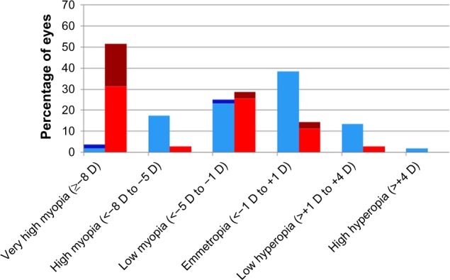
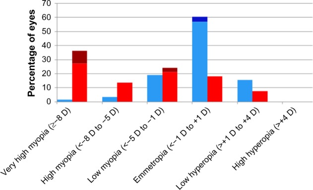
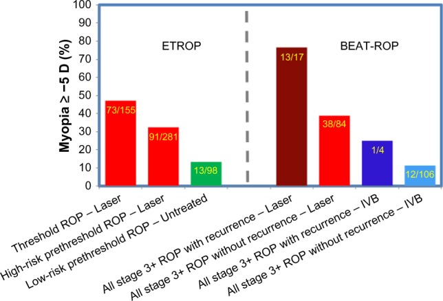
To examine the effect of anti-vascular endothelial growth factor (anti-VEGF) agents on refractive error in the setting of retinopathy of prematurity (ROP) through a review of the literature, a PubMed search was performed of appropriate search terms, and the results of all relevant studies were extracted and compiled. Eleven relevant articles were identified in the literature, totaling 466 eyes, treated with varied anti-VEGF agents (bevacizumab, ranibizumab, and aflibercept) with mean spherical equivalent refractions ranging from +0.75 D to -3.57 D, with prevalence of high myopia ranging from 0 to 35%. Anti-VEGF monotherapy for ROP leads to low levels of myopia, and there may be a differential effect of specific anti-VEGF agents utilized on refractive outcomes.

Keywords: ROP; aflibercept; bevacizumab; myopia; ranibizumab; refraction; retinopathy of prematurity.


Conflict of interest statement

Disclosure The authors report no conflicts of interests in this work.

Figures

Figure 1
Figure 1
Zone I distribution of refractive error by treatment modality. Notes: Distribution of spherical equivalent refractive error at age 2.5 years in eyes that received treatment for stage 3+ retinopathy of prematurity or aggressive posterior retinopathy of prematurity in the Bevacizumab Eliminates the Angiogenic Threat for Retinopathy of Prematurity clinical trial. Data presented according to treatment modality: red, laser without recurrence; brown, laser with recurrence; light blue, intravitreal bevacizumab without recurrence; dark blue, intravitreal bevacizumab with recurrence. Reproduced with permission from JAMA Ophthalmol. 2014;132(11):1327–1333. Copyright ©2014 American Medical Association. All rights reserved.
Figure 2
Figure 2
Zone II posterior distribution of refractive error by treatment modality. Notes: Distribution of spherical equivalent refractive error at age 2.5 years in eyes that received treatment for stage 3+ retinopathy of prematurity or aggressive posterior retinopathy of prematurity in the Bevacizumab Eliminates the Angiogenic Threat for Retinopathy of Prematurity clinical trial. Data presented according to treatment modality: red, laser without recurrence; brown, laser with recurrence; light blue, intravitreal bevacizumab without recurrence; dark blue, intravitreal bevacizumab with recurrence. Reproduced with permission from JAMA Ophthalmol. 2014;132(11):1327–1333. Copyright © 2014 American Medical Association. All rights reserved.
Figure 3
Figure 3
Percentage of eyes ≥−5 D in ETROP and BEAT-ROP by ROP severity at treatment. Notes: Percentage of eyes with myopia ≥−5 D in the ETROP and BEAT-ROP clinical trials at ages 3 and 2.5 years, respectively. Data presented according to ROP severity at treatment (threshold, high-risk prethreshold, low-risk prethreshold, and all stage 3+) by treatment modality utilized (laser and IVB) in the respective clinical trials. Data from ETROP, and BEAT-ROP. Abbreviations: ROP, retinopathy of prematurity; IVB, intravitreal bevacizumab.

References

    1. Geloneck MM, Chuang AZ, Clark WL, et al. Refractive outcomes following bevacizumab monotherapy compared with conventional laser treatment: a randomized clinical trial. JAMA Ophthalmol. 2014;132(11):1327–1333. - PubMed
    1. Quinn GE, Dobson V, Kivlin J, et al. Prevalence of myopia between 3 months and 5½ years in preterm infants with and without retinopathy of prematurity. Ophthalmology. 1998;105(7):1292–1300. - PubMed
    1. Quinn GE, Dobson V, Davitt BV, et al. Progression of myopia and high myopia in the Early Treatment for Retinopathy of Prematurity study: findings to 3 years of age. Ophthalmology. 2008;115(6):1058–1064.e1. - PubMed
    1. Quinn GE, Dobson V, Davitt BV, et al. Progression of myopia and high myopia in the Early Treatment for Retinopathy of Prematurity study: findings at 4 to 6 years of age. J AAPOS. 2013;17(2):124–128. - PMC - PubMed
    1. Nissenkorn I, Yassur Y, Mashkowski D, Sherf I, Ben-Sira I. Myopia in premature babies with and without retinopathy of prematurity. Br J Ophthalmol. 1983;67(3):170–173. - PMC - PubMed

LinkOut - more resources