

Bullous lichen planus - a review
- PMID: 28539981
- PMCID: PMC5439688
- DOI: 10.3315/jdcr.2017.1239
Bullous lichen planus - a review
Abstract
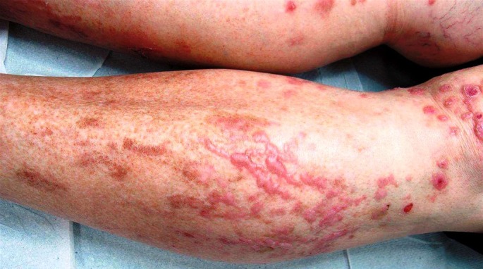
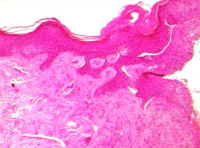
Bullous lichen planus is a rare variant of lichen planus. It is characterized by vesicles or bullae, which usually develop in the context of pre-existing LP lesions. It is often misdiagnosed and should be differentiated from other subepidermal bullous diseases especially lichen planus pemphigoides. The diagnosis is based on clinical suspicion and is confirmed by histopathology and immunofluoresence. The clinical features of bullous lichen planus include typical lichen planus lesions, accompanied by the formation of bullae on the affected or perilesional skin. This is evident on histology, with alteration of the dermo-epidermal junction and intrabasal bullae as a consequence of extensive inflammation. The histologic features in conjunction with the negative immunofluoresence indicate that bullous lichen planus is a form of "hyper-reactive lichen planus" rather than a distinct entity. There is no standard treatment of bullous lichen planus. Topical and systemic corticosteroids, dapsone and acitretin have been described as effective choices.
Keywords: bullous lichen planus; lichen planus; lichen planus pemphigoides; review.
Figures
References
-
- Pinkus H. Lichenoid tissue reactions. A speculative review of the clinical spectrum of epidermal basal cell damage with special reference to erythema dyschromicum perstans. Arch Dermatol. 1973;107:840–846. - PubMed
-
- Huang C, Chen S, Liu Z, Tao J, Wang C, Zhou Y. Familial bullous lichen planus (FBLP): Pedigree analysis and clinical characteristics. J Cutan Med Surg. 2005;9:217–222. - PubMed
-
- Huang C, Yan X, Yang L, Zhang J, Tian J, Li J, Wang C, Tu Y. A retrospective and comparative study of familial and non-familial bullous lichen planus. Huazhong Univ Sci Technolog Med Sci. 2007;27:336–338. - PubMed
-
- Zhou XJ, Sugerman PB, Savage NW, Walsh LJ, Seymour GJ. Intra-epithelial CD8+ T cells and basement membrane disruption in oral lichen planus. J Oral Pathol Med. 2002;31:23–27. - PubMed
-
- Scully C, Beyli M, Ferreiro MC, Ficarra G, Gill Y, Griffiths M, Holmstrup P, Mutlu S, Porter S, Wray D. Update on oral lichen planus: etiopathogenesis and management. Crit Rev Oral Biol Med. 1998;9:86–122. - PubMed
Publication types
LinkOut - more resources
Full Text Sources
Other Literature Sources