

Outgrowing endothelial and smooth muscle cells for tissue engineering approaches
- PMID: 28540031
- PMCID: PMC5433677
- DOI: 10.1177/2041731417698852
Outgrowing endothelial and smooth muscle cells for tissue engineering approaches
Abstract
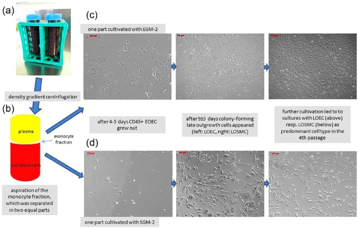
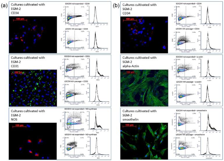
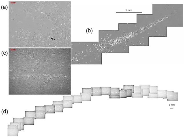
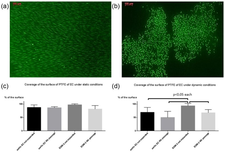
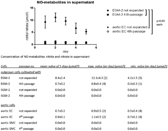
In recent years, circulating progenitors of endothelial cells and smooth muscle cells were identified in the peripheral blood. In our study, we evaluated the utilization of both cell types isolated and differentiated from peripheral porcine blood in terms for their use for tissue engineering purposes. By means of density gradient centrifugation, the monocyte fraction from porcine blood was separated, split, and cultivated with specific culture media with either endothelial cell growth medium-2 or smooth muscle cell growth medium-2 for the differentiation of endothelial cells or smooth muscle cells. Obtained cells were characterized at an early stage of cultivation before the first passage and a late stage (fourth passage) on the basis of the expression of the antigens CD31, CD34, CD45, nitric oxide synthase, and the contractile filaments smooth-muscle alpha-actin (sm-alpha-actin) and smoothelin. Functional characterization was done based on the secretion of nitric oxide, the formation of a coherent monolayer on polytetrafluoroethylene, and capillary sprouting. During cultivation in both endothelial cell growth medium-2 and smooth muscle cell growth medium-2, substantially two types of cells grew out: early outgrown CD45-positive cells, which disappeared during further cultivation, and in 85% (n = 17/20) of cultures cultivated with endothelial cell growth medium-2 colony-forming late outgrowth endothelial cells. During cultivation with smooth muscle cell growth medium-2 in 80% (n = 16/20) of isolations colony-forming late outgrowth smooth muscle cells entered the stage. Cultivation with either endothelial cell growth medium-2 or smooth muscle cell growth medium-2 had selective effect on the late outgrown cells to that effect that the number of CD31-positive cells increased from 34.8% ± 13% to 83.9% ± 8% in cultures cultivated with endothelial cell growth medium-2 and the number of sm-α-actin+ cells increased from 52.6% ± 18% to 88% ± 5% in cultures cultivated with smooth muscle cell growth medium-2, respectively. Functional analyses revealed significantly higher levels of nitric oxide secretion, endothelialization capacity, and capillary formation in not expanded cultures cultivated with endothelial cell growth medium-2 in comparison to later stages of cultivation and mature aortic cells. Blood seems to be a reliable and feasible source for the isolation of both endothelial and smooth muscle cells for application in tissue engineering approaches. Whereas, early co-cultures of early and late outgrowth cells provide functional advantages, the differentiation of cells can be directed selectively by the used culture medium for the expansion of highly proliferative late outgrowth endothelial cells and late outgrowth smooth muscle cells, respectively.
Keywords: Circulating progenitor cells; early outgrowth endothelial cells; endothelial progenitor cells; late outgrowth endothelial cells; late outgrowth smooth muscle cells; tissue engineering.
Conflict of interest statement
Declaration of conflicting interests: The authors state that there are commercial associations that might create a conflict of interest including financial interests in connection with this manuscript.
Figures
Similar articles
-
[BIOLOGICAL FEATURES AND IDENTIFICATION OF ENDOTHELIAL PROGENITOR CELLS FROM PERIPHERAL BLOOD].Zhongguo Xiu Fu Chong Jian Wai Ke Za Zhi. 2015 Jul;29(7):870-7. Zhongguo Xiu Fu Chong Jian Wai Ke Za Zhi. 2015. PMID: 26540983 Chinese.
-
Vascular progenitor cells isolated from human embryonic stem cells give rise to endothelial and smooth muscle like cells and form vascular networks in vivo.Circ Res. 2007 Aug 3;101(3):286-94. doi: 10.1161/CIRCRESAHA.107.150201. Epub 2007 Jun 14. Circ Res. 2007. PMID: 17569886
-
Smooth muscle progenitor cells in human blood.Circulation. 2002 Sep 3;106(10):1199-204. doi: 10.1161/01.cir.0000031525.61826.a8. Circulation. 2002. PMID: 12208793
-
Clinical Application of Endothelial Progenitor Cell: Are We Ready?Acta Cardiol Sin. 2013 Nov;29(6):479-87. Acta Cardiol Sin. 2013. PMID: 27122748 Free PMC article. Review.
-
Cooperation of liver cells in health and disease.Adv Anat Embryol Cell Biol. 2001;161:III-XIII, 1-151. doi: 10.1007/978-3-642-56553-3. Adv Anat Embryol Cell Biol. 2001. PMID: 11729749 Review.
Cited by
-
A bioassay system of autologous human endothelial, smooth muscle cells, and leukocytes for use in drug discovery, phenotyping, and tissue engineering.FASEB J. 2020 Jan;34(1):1745-1754. doi: 10.1096/fj.201901379RR. Epub 2019 Dec 5. FASEB J. 2020. PMID: 31914612 Free PMC article.
-
3D Tissue-Engineered Vascular Drug Screening Platforms: Promise and Considerations.Front Cardiovasc Med. 2022 Mar 4;9:847554. doi: 10.3389/fcvm.2022.847554. eCollection 2022. Front Cardiovasc Med. 2022. PMID: 35310996 Free PMC article. Review.
-
Development and Preliminary Testing of Porcine Blood-Derived Endothelial-like Cells for Vascular Tissue Engineering Applications: Protocol Optimisation and Seeding of Decellularised Human Saphenous Veins.Int J Mol Sci. 2022 Jun 14;23(12):6633. doi: 10.3390/ijms23126633. Int J Mol Sci. 2022. PMID: 35743073 Free PMC article.
-
Novel Cell-Based and Tissue Engineering Approaches for Induction of Angiogenesis as an Alternative Therapy for Diabetic Retinopathy.Int J Mol Sci. 2020 May 15;21(10):3496. doi: 10.3390/ijms21103496. Int J Mol Sci. 2020. PMID: 32429094 Free PMC article. Review.
-
Developing human upper, lower, and deep lung airway models: Combining different scaffolds and developing complex co-cultures.J Tissue Eng. 2025 Jan 30;16:20417314241299076. doi: 10.1177/20417314241299076. eCollection 2025 Jan-Dec. J Tissue Eng. 2025. PMID: 39885949 Free PMC article.
References
-
- Simper D, Stalboerger PG, Panetta CJ, et al. Smooth muscle progenitor cells in human blood. Circulation 2002; 106(10): 1199–1204. - PubMed
-
- Asahara T, Murohara T, Sullivan A, et al. Isolation of putative progenitor endothelial cells for angiogenesis. Science 1997; 275(5302): 964–967. - PubMed
-
- Guan XM, Cheng M, Li H, et al. Biological properties of bone marrow-derived early and late endothelial progenitor cells in different culture media. Mol Med Rep 2013; 8(6): 1722–1728. - PubMed
-
- Rehman J, Li J, Orschell CM, et al. Peripheral blood “endothelial progenitor cells” are derived from monocyte/macrophages and secrete angiogenic growth factors. Circulation 2003; 107(8): 1164–1169. - PubMed
-
- Rehman J, Li J, Parvathaneni L, et al. Exercise acutely increases circulating endothelial progenitor cells and monocyte-/macrophage-derived angiogenic cells. J Am Coll Cardiol 2004; 43(12): 2314–2318. - PubMed
LinkOut - more resources
Full Text Sources
Other Literature Sources
Research Materials
Miscellaneous
