

Review
doi: 10.21037/jtd.2017.03.141.
Integrate imaging approach for minimally invasive and robotic procedures
Affiliations
- PMID: 28540069
- PMCID: PMC5422659
- DOI: 10.21037/jtd.2017.03.141
Item in Clipboard
Review
Integrate imaging approach for minimally invasive and robotic procedures
J Thorac Dis.
2017 Apr.
Abstract
Over the past two decades, robotic and minimally invasive cardiac surgery has been continuously refined and is currently an alternative to traditional open-heart surgery for some patients. The parallel evolution of imaging modalities has made robotic surgery safer and more efficient. Here, we review the pre- and post-operative use of computed tomography (CT) in minimally invasive and robotic cardiac procedures.
Keywords: Cardiac surgical procedures; X-ray computed tomography; minimally invasive surgical procedures; mitral valve annuloplasty; vascular graft occlusion.
Conflict of interest statement
Conflicts of Interest: The authors have no conflicts of interest to declare.
Figures
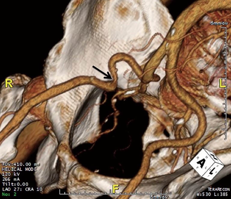
Three-dimensional (3D) volume rendered image obtained from a computed tomography (CT) scan showing a tortuous right external iliac artery with a narrowing (indicated by the arrow). In contrast, the left external iliac artery is normal.
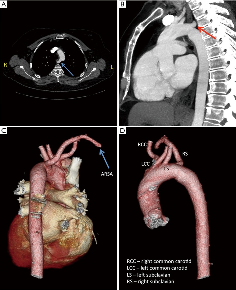
Computed tomography (CT) scans demonstrating an aberrant right subclavian artery (ARSA)—instead of taking origin from the brachiocephalic artery the ARSA is the last branch off the aortic arch and loops around to reach the right side. (A,B) ARSA is indicated by the arrow; (C,D) three-dimensional (3D) volume rendered images obtained from a CT scan.
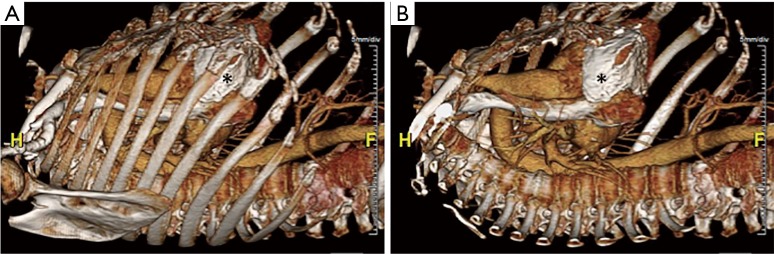
Three-dimensional (3D) volume rendered images obtained from a computed tomography (CT) scan with ribs present (A) and digitally removed (B). Areas of pericardial calcifications can be easily seen and are indicated by asterisks.
Similar articles
-
Three-Dimensional Printing of Life-Like Models for Simulation and Training of Minimally Invasive Cardiac Surgery.Innovations (Phila). 2017 Nov/Dec;12(6):459-465. doi: 10.1097/IMI.0000000000000423. Innovations (Phila). 2017. PMID: 29232300 Free PMC article.
-
Preoperative multidetector computed tomograpy angiography for planning of minimally invasive robotic mitral valve surgery: impact on decision making.J Thorac Cardiovasc Surg. 2013 Aug;146(2):262-8.e1. doi: 10.1016/j.jtcvs.2012.06.052. Epub 2012 Jul 25. J Thorac Cardiovasc Surg. 2013. PMID: 22841167
-
Minimally invasive robotic mitral valve surgery.Expert Rev Med Devices. 2011 Jan;8(1):115-20. doi: 10.1586/erd.10.66. Expert Rev Med Devices. 2011. PMID: 21158546 Review.
-
Robotic minimally invasive mitral valve reconstruction yields less blood product transfusion and shorter length of stay.Surgery. 2006 Aug;140(2):263-7. doi: 10.1016/j.surg.2006.05.003. Surgery. 2006. PMID: 16904978
-
Robotics in cardiac surgery.Scand J Surg. 2009;98(2):120-4. doi: 10.1177/145749690909800207. Scand J Surg. 2009. PMID: 19799049 Review.
Cited by
-
State-of-the-Art Review: Technical and Imaging Considerations in Novel Transapical and Port-Access Mitral Valve Chordal Repair for Degenerative Mitral Regurgitation.Front Cardiovasc Med. 2022 Apr 12;9:850700. doi: 10.3389/fcvm.2022.850700. eCollection 2022. Front Cardiovasc Med. 2022. PMID: 35497995 Free PMC article. Review.
-
Complications and their management in robotic mitral valve surgery from the surgical assistant's perspective.Ann Cardiothorac Surg. 2022 Sep;11(5):510-524. doi: 10.21037/acs-2022-rmvs-15. Ann Cardiothorac Surg. 2022. PMID: 36237594 Free PMC article. Review.
References
Publication types
LinkOut - more resources
Full Text Sources
Other Literature Sources
Miscellaneous