

Intra-Articular Osteoid Osteoma as a Cause of Anteromedial Knee Pain
- PMID: 28540096
- PMCID: PMC5433411
- DOI: 10.1155/2017/5846368
Intra-Articular Osteoid Osteoma as a Cause of Anteromedial Knee Pain
Abstract
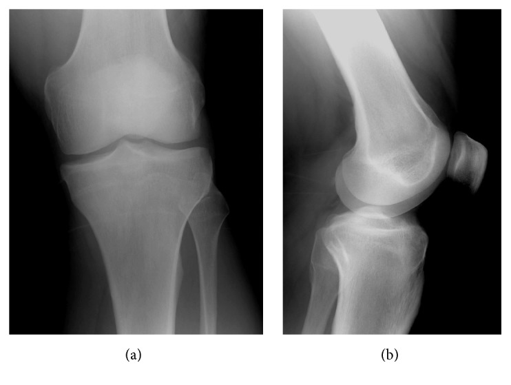
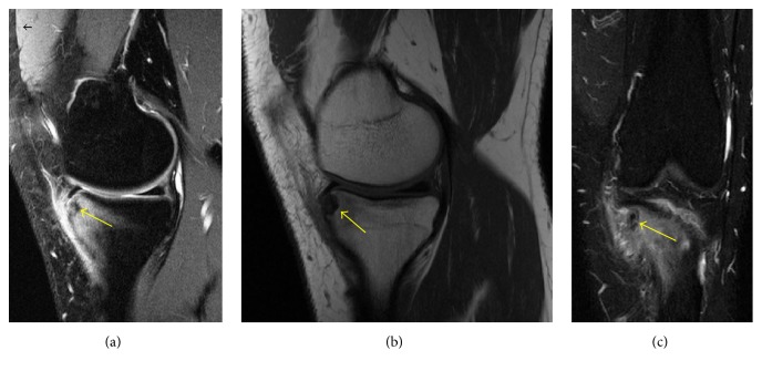
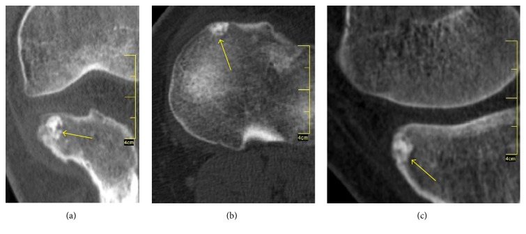
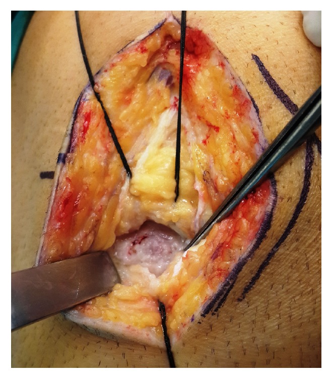
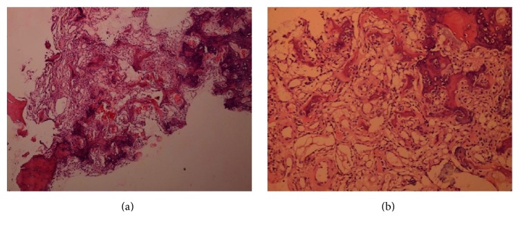
A 32-year-old male patient presented to our clinic with chronic left knee pain that was ongoing for about 1.5 years. The patient visited several times our clinic and the other clinics; conservative treatment (including rest, knee brace, and ice application with NSAIDs) was recommended by various different doctors. The anamnesis, physical examination, and plain radiography were nonspecific. Early MRI findings mislead us to believe it is bone marrow edema. One and half years with noneffective treatment, the knee pain persisted. At the latest visit intra-articular osteoid osteoma was suspected and the knee MRI with CT was employed. Even though the diagnosis of intra-articular osteoid osteoma often presents a challenge for the surgeons, with a present awareness of intra-articular osteoid osteomas which lack the characteristic sclerotic lesions and nidus on plain X-rays and the aid of multislice CT, a correct diagnosis which warrants proper treatment can be achieved. The possibility of osteoid osteomas, especially in young adults with persistent knee pain with unknown reasons that show normal plain radiographs results, must not be overlooked. The treatment method of these lesions should be customized depending on the location of the lesion, experience of the surgeon, and cost of method.
Figures
References
-
- Jaffe H. L. Osteoid Osteoma, a benign osteoblastic tumor composed of osteoid and atypical bone. Archieves of Surgery. 1935;31:708–723.
Publication types
LinkOut - more resources
Full Text Sources
Other Literature Sources
