Scalpel Depigmentation and Surgical Crown Lengthening to Improve Anterior Gingival Esthetics
- PMID: 28540108
- PMCID: PMC5423340
- DOI: 10.4103/amhsr.amhsr_442_12
Scalpel Depigmentation and Surgical Crown Lengthening to Improve Anterior Gingival Esthetics
Abstract
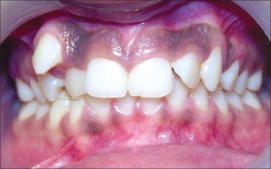
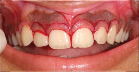
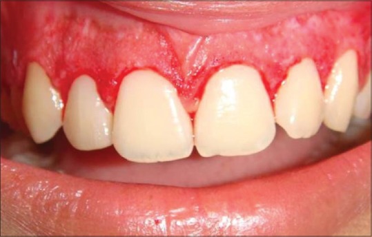
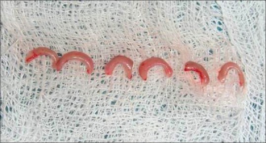
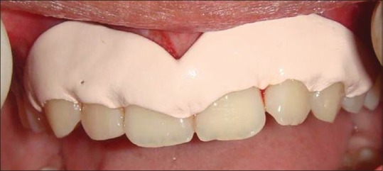
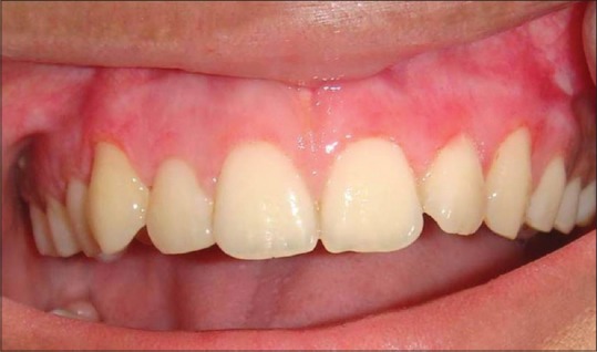
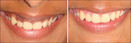
A smile is a mesmerizing expression of joy, success, courtesy and reveals self-confidence. The harmony of the smile is determined not only by the shape, position and the color of teeth, but also by the gingival architecture. Gingival tissues form an important part of what we can consider to be a pleasing smile. The elegance of this pleasant smile could be, affected by highly pigmented gingiva. This gingival pigmentation occurs as a result of melanin granules, which are produced by melanoblast. Although melanin pigmentation does not present a medical problem, demand for cosmetic therapy is commonly sort by people with moderately pigmented gingiva. This case report highlights, a case of excessive gingival display and hyperpigmentation treated with anterior crown lengthening and gingival depigmentation using the scalpel technique.
Keywords: Depigmentation; Gingiva; Melanin; Scalpel technique.
Conflict of interest statement
There are no conflicts of interest.
Figures
Similar articles
-
Comparison between scalpel technique and electrosurgery for depigmentation: A case series.J Indian Soc Periodontol. 2011 Oct;15(4):402-5. doi: 10.4103/0972-124X.92580. J Indian Soc Periodontol. 2011. PMID: 22368368 Free PMC article.
-
Anterior esthetic gingival depigmentation and crown lengthening: report of a case.J Contemp Dent Pract. 2005 Aug 15;6(3):139-47. J Contemp Dent Pract. 2005. PMID: 16127483
-
Treatment of gingival pigmentation: a case series.Indian J Dent Res. 2005 Oct-Dec;16(4):171-6. doi: 10.4103/0970-9290.29901. Indian J Dent Res. 2005. PMID: 16761712
-
Achieving better esthetics by gingival de-pigmentation: report of three cases with a review of the literature.J Mich Dent Assoc. 2013 Feb;95(2):52-8, 78. J Mich Dent Assoc. 2013. PMID: 23556274 Review.
-
Esthetic crown lengthening for maxillary anterior teeth.Compend Contin Educ Dent. 1997 Aug;18(8):807-12, 814-6, 818-9; quiz 820. Compend Contin Educ Dent. 1997. PMID: 9533339 Review.
References
-
- Liebart MF, Fouque-Deruelle C, Santini A, Dillier FL, Monnet-Corti V, Glise JM, et al. Smile line and periodontium visibility. Periodontol. 2000;1:17–25.
-
- Granstein RD, Sober AJ. Drug- and heavy metal – Induced hyperpigmentation. J Am Acad Dermatol. 1981;5:1–18. - PubMed
-
- Coslet JG, Vanarsdall R, Weisgold A. Diagnosis and classification of delayed passive eruption of the dentogingival junction in the adult. Alpha Omegan. 1977;70:24–8. - PubMed
-
- Hürzeler MB, Weng D. Functional and esthetic outcome enhancement of periodontal surgery by application of plastic surgery principles. Int J Periodontics Restorative Dent. 1999;19:36–43. - PubMed
-
- Dummett CO. Oral pigmentation. First symposium of oral pigmentation. J Periodontol. 1960;31:356–60.
Publication types
LinkOut - more resources
Full Text Sources
Other Literature Sources