

A case of a cerebral cavernous malformation of the third ventricle that caused the syndrome of inappropriate secretion of antidiuretic hormone
- PMID: 28540119
- PMCID: PMC5421201
- DOI: 10.4103/sni.sni_1_17
A case of a cerebral cavernous malformation of the third ventricle that caused the syndrome of inappropriate secretion of antidiuretic hormone
Abstract
Background: Cerebral cavernous malformations (CCMs, also known as cavernous hemanigiomas) of the third ventricle are uncommon. Here, we present a rare case of a CCM that caused the syndrome of inappropriate secretion of antidiuretic hormone (SIADH).
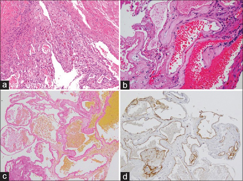
Case description: A 68-year-old man presented with acute-onset cognitive and memory disturbance. Endocrinological examinations revealed hyponatremia due to SIADH. Computed tomography indicated a high-density mass in the third ventricle that caused left unilateral hydrocephalus due to obstruction of the foramen Monroe. On magnetic resonance imaging, the mass showed high intensity in both T1 and T2-weighted images and low intensity in susceptibility-weighted images, suggesting subacute intralesional hemorrhage. We completely excised the mass via a basal interhemispheric translamina terminalis approach. Intraoperatively, the mass adhered tightly to the left hypothalamus, which was supposed to the origin and was well circumscribed from the surroundings. The histopathological diagnosis was CCM, and his SIADH improved after the operation.
Conclusion: We presented a rare case of a CCM in the third ventricle that caused SIADH, which improved after complete excision of the mass via a basal interhemispheric translamina terminalis approach.
Keywords: Cavernous hemangioma; SIADH; cerebral cavernous malformation; third ventricle.
Conflict of interest statement
There are no conflicts of interest.
Figures


Similar articles
-
A large cavernous malformation of the third ventricle floor: A case report.Neurol Neurochir Pol. 2015;49(6):446-50. doi: 10.1016/j.pjnns.2015.08.004. Epub 2015 Sep 4. Neurol Neurochir Pol. 2015. PMID: 26652881
-
Surgical management of cavernous malformations of the third ventricle.J Neurosurg. 1994 Jan;80(1):64-72. doi: 10.3171/jns.1994.80.1.0064. J Neurosurg. 1994. PMID: 8271024 Review.
-
Interhemispheric, Translamina Terminalis Approach for the Resection of Suprasellar Cavernous Malformation.J Neurol Surg B Skull Base. 2018 Apr;79(Suppl 3):S278. doi: 10.1055/s-0038-1624588. Epub 2018 Mar 7. J Neurol Surg B Skull Base. 2018. PMID: 29588900 Free PMC article.
-
Endoscopic Endonasal Resection of a Cavernous Malformation of the Third Ventricle: Case Report and Literature Review.J Neurol Surg A Cent Eur Neurosurg. 2024 Mar;85(2):221-226. doi: 10.1055/s-0041-1741070. Epub 2022 May 29. J Neurol Surg A Cent Eur Neurosurg. 2024. PMID: 35644135 Review.
-
Rapid deterioration of primary fourth ventricular outlet obstruction resulting in syndrome of inappropriate antidiuretic hormone secretion.Pediatr Int. 2014 Aug;56(4):e30-2. doi: 10.1111/ped.12387. Pediatr Int. 2014. PMID: 25252067
Cited by
-
Hypothalamic Cavernous Malformation: Surgical Technique and Literature Review.Cureus. 2022 Jan 23;14(1):e21511. doi: 10.7759/cureus.21511. eCollection 2022 Jan. Cureus. 2022. PMID: 35223287 Free PMC article.
References
-
- Beier AD, Cheshier SH, Chakraborty A, Dirks P. Suprasellar arachnoid cyst resulting in the syndrome of inappropriate antidiuretic hormone secretion. J Neurosurg Pediatr. 2010;6:486–8. - PubMed
-
- Edate S, Albanese A. Management of Electrolyte and Fluid Disorders after Brain Surgery for Pituitary/Suprasellar Tumours. Horm Res Paediatr. 2015;83:293–301. - PubMed
-
- Faropoulos K, Panagiotopoulos V, Partheni M, Tzortzidis F, Konstantinou D. Therapeutic management of intraventricular cavernoma: Case series and review of the literature. J Neurol Surg. 2015;76:233–9. - PubMed
-
- Gross BA, Ning Lin, Rose Du, Arthur L. Day. The natural history of intracranial cavernous malformations. Neurosurg Focus. 2011;30:E24. - PubMed
-
- Katayama Y, Tsubokawa T, Maeda T, Yamamoto T. Surgical management of cavernous malformations of the third ventricle. J Neurosurg. 1994;80:64–72. - PubMed
Publication types
LinkOut - more resources
Full Text Sources
Other Literature Sources