Corpora amylacea mimicking low-grade glioma and manifesting as a seizure: Case report
- PMID: 28540130
- PMCID: PMC5421196
- DOI: 10.4103/sni.sni_423_16
Corpora amylacea mimicking low-grade glioma and manifesting as a seizure: Case report
Abstract
Background: Corpora amylacea (CA) are accumulations of polyglucosan bodies typically found in astrocytic foot processes, and rarely, can mimic neoplasm. CA accumulation has also been associated with seizure disorders. We report the first case of a histologically confirmed intracranial, intraparenchymal CA lesion mimicking a low-grade glioma and manifesting as a seizure.
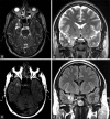
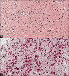
Case description: A 43-year-old man presented after a general tonic-clonic (GTC) seizure. Brain magnetic resonance imaging (MRI) revealed a small lesion in the right mesial temporal lobe with radiologic features of a low-grade glioma. The patient underwent a right pteronial craniotomy for resection of the lesion. Histology demonstrated abundant polyglucosan bodies without neoplastic features. The patient tolerated the procedure well, was free from seizures without antiepileptic drugs at 2-week follow-up, and is undergoing serial surveillance.
Conclusion: The clinical manifestation of CA as a seizure in the context of an identified brain mass is extraordinarily rare. Nevertheless, CA should be considered in the differential diagnosis for patients with seizures and a radiologically identifiable low-grade lesion. Symptomatic CA lesions Mimicking a low-grade glioma should be surgically pursued with a goal of safe, maximal resection to confirm the diagnosis and to provide the patient with prognosis, which can significantly impact patient quality of life.
Keywords: Case reports; corpora amylacea; glioma; pathology; seizures.
Conflict of interest statement
There are no conflicts of interest.
Figures


Similar articles
-
Diffuse low grade glioma after the 2016 WHO update, seizure characteristics, imaging correlates and outcomes.Clin Neurol Neurosurg. 2018 Dec;175:9-15. doi: 10.1016/j.clineuro.2018.10.001. Epub 2018 Oct 2. Clin Neurol Neurosurg. 2018. PMID: 30292978
-
Awake Craniotomy with Noninvasive Brain Mapping by 3-Tesla Functional Magnetic Resonance Imaging for Excision of Low-grade Glioma: A Case of a Young Patient from Pakistan.Asian J Neurosurg. 2018 Apr-Jun;13(2):471-474. doi: 10.4103/ajns.AJNS_144_16. Asian J Neurosurg. 2018. PMID: 29682064 Free PMC article.
-
An extent of resection threshold for seizure freedom in patients with low-grade gliomas.J Neurosurg. 2018 Apr;128(4):1084-1090. doi: 10.3171/2016.12.JNS161682. Epub 2017 May 26. J Neurosurg. 2018. PMID: 28548599
-
Angiocentric Glioma in an Elderly Patient: Case Report and Review of the Literature.World Neurosurg. 2017 Jan;97:755.e5-755.e10. doi: 10.1016/j.wneu.2016.10.034. Epub 2016 Oct 15. World Neurosurg. 2017. PMID: 27756673 Review.
-
[Developmental venous anomaly presenting as a brain parenchymal abnormality mimicking a brain tumor:a report of 3 cases and review of literature].Brain Nerve. 2010 Dec;62(12):1331-6. Brain Nerve. 2010. PMID: 21139186 Review. Japanese.
Cited by
-
Distribution of corpora amylacea in the human midbrain: using synchrotron radiation phase-contrast microtomography, high-field magnetic resonance imaging, and histology.Front Neurosci. 2023 Oct 4;17:1236876. doi: 10.3389/fnins.2023.1236876. eCollection 2023. Front Neurosci. 2023. PMID: 37869518 Free PMC article.
-
Wasteosomes (corpora amylacea) as a hallmark of chronic glymphatic insufficiency.Proc Natl Acad Sci U S A. 2022 Nov 29;119(48):e2211326119. doi: 10.1073/pnas.2211326119. Epub 2022 Nov 21. Proc Natl Acad Sci U S A. 2022. PMID: 36409907 Free PMC article.
-
Case report: The origin of transmantle-like features.Front Radiol. 2022 Jul 29;2:927764. doi: 10.3389/fradi.2022.927764. eCollection 2022. Front Radiol. 2022. PMID: 37492659 Free PMC article.
References
-
- Abel TJ, Hebb AO, Keene CD, Born DE, Silbergeld DL. Parahippocampal corpora amylacea: Case report. Neurosurgery. 2010;66:E1206–7. - PubMed
-
- Bigio EH, Weiner MF, Bonte FJ, White CL. Familial dementia due to adult polyglucosan body disease. Clin Neuropathol. 1997;16:227–34. - PubMed
-
- Busard HL, Gabreels-Festen AA, Renier WO, Gabreels FJ, Joosten EM, van ‘t Hof MA, et al. Adult polyglucosan body disease: The diagnostic value of axilla skin biopsy. Ann Neurol. 1991;29:448–51. - PubMed
-
- Cavanagh JB. Corpora-amylacea and the family of polyglucosan diseases. Brain Res Brain Res Rev. 1999;29:265–95. - PubMed
-
- Chen W, Silverman DH, Delaloye S, Czernin J, Kamdar N, Pope W, et al. 18F-FDOPA PET imaging of brain tumors: Comparison study with 18F-FDG PET and evaluation of diagnostic accuracy. J Nucl Med. 2006;47:904–11. - PubMed
Publication types
LinkOut - more resources
Full Text Sources
Other Literature Sources
