Review
. 2017 Apr 26:8:65.
doi: 10.4103/sni.sni_441_16. eCollection 2017.

Parkinson's disease: Microglial/macrophage-induced immunoexcitotoxicity as a central mechanism of neurodegeneration

Affiliations
Review

Parkinson's disease: Microglial/macrophage-induced immunoexcitotoxicity as a central mechanism of neurodegeneration

Russell L Blaylock. Surg Neurol Int. .

Abstract

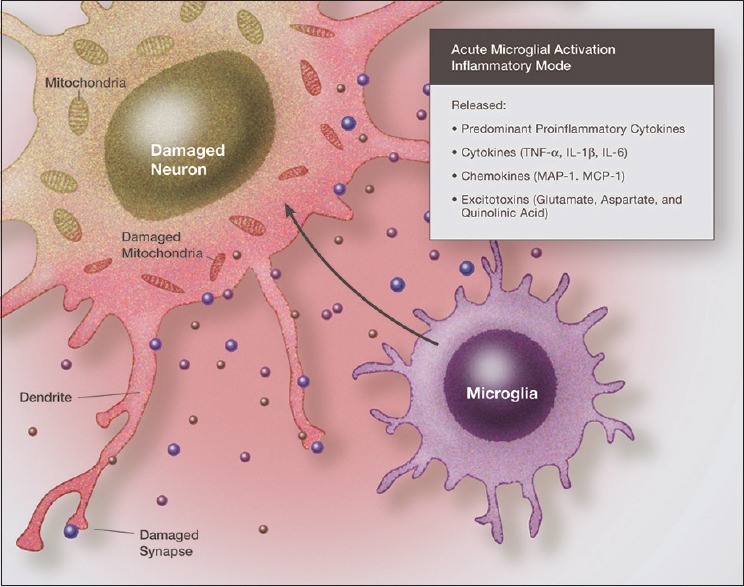
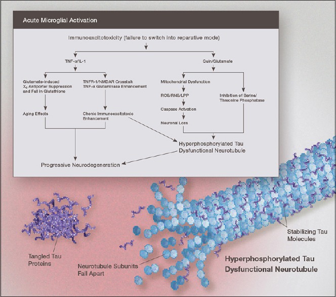
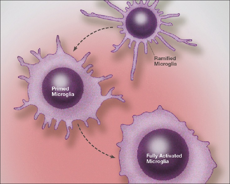
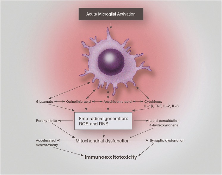
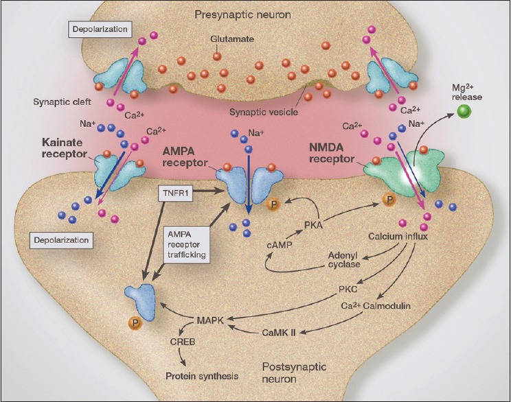
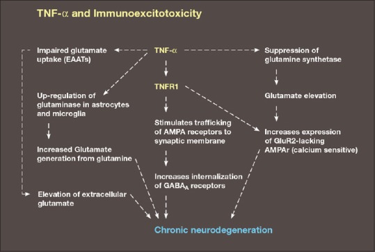
Parkinson's disease is one of the several neurodegenerative disorders that affects aging individuals, with approximately 1% of those over the age of 60 years developing the disorder in their lifetime. The disease has the characteristics of a progressive disorder in most people, with a common pattern of pathological change occurring in the nervous system that extends beyond the classical striatal degeneration of dopaminergic neurons. Earlier studies concluded that the disease was a disorder of alpha-synuclein, with the formation of aggregates of abnormal alpha-synuclein being characteristic. More recent studies have concluded that inflammation plays a central role in the disorder and that the characteristic findings can be accounted for by either mutation or oxidative damage to alpha-synuclein, with resulting immune reactions from surrounding microglia, astrocytes, and macrophages. What has been all but ignored in most of these studies is the role played by excitotoxicity and that the two processes are intimately linked, with inflammation triggered cell signaling enhancing the excitotoxic cascade. Further, there is growing evidence that it is the excitotoxic reactions that actually cause the neurodegeneration. I have coined the name immunoexcitotoxicity to describe this link between inflammation and excitotoxicity. It appears that the two processes are rarely, if ever, separated in neurodegenerative diseases.

Keywords: Alzheimer’s; Parkinson's disease; immunoexcitotoxicity; microglial activation; microglial priming; neurodegenerative diseases.


Figures

Figure 1
Figure 1
Illustration demonstrating the neurotoxic effects of proinflammatory cytokines and chemokines acting in synergy with excitatory amino acids. Immunoexcitoxicity results in damage to neuronal cell membranes, mitochondria, DNA, as well as dendrites and synapses mainly by the excitotoxic mechanisms
Figure 2
Figure 2
Illustration of the mechanism linking immunoexcitotoxicity with increased deposition of hyperphosphorylated tau in the chronically inflamed brain. Once the microglia fail to switch to the reparative or resting mode, elevated levels of IL-1ß, TNF-, and other proinflammatory cytokines and chemokines, act to enhance excitotoxicity by a number of mechanisms. High levels of glutamate and nitric oxide suppress mitochondrial energy generation. Both elevated QUIN and glutamate increase levels of oxidized and nitrated alpha-synuclein
Figure 3
Figure 3
Illustration of microglia priming/activation transition states beginning from a resting (ramified) state. Recent studies indicate that microglia can assume a number of activation states, such as predominately phagocytic, predominately neuroprotective or predominately neurodestructive. In the primed state the mRNA for cytokines, chemokines and other reactive molecules are upregulated but active proteins are not released
Figure 4
Figure 4
Illustration of the neurotoxic factors released from an activated microglia, demonstrating the interaction of proinflammatory cytokines, prostaglandins and excitatory amino acids. Of particular importance is the effect on mitochondrial function, which when depressed enhances excitotoxic sensitivity as well as reactive oxygen species generation
Figure 5
Figure 5
Illustration of glutamatergic synapse demonstrating rapid AMPA receptor trafficking from the endoplasmic reticulum, which is driven by activation of TNFR1. Crosstalk between the AMPA receptor and TNFR1 increase synaptic insertion of GluR2-lacking (calcium permeable) AMPA receptors, thus increasing synaptic glutamate-related sensitivity (excitotoxicity). TNFR1 activation also increases GABA receptor endocytosis, which increases synaptic sensitivity to excitotoxicity even further
Figure 6
Figure 6
Diagram demonstrating a number of the major mechanisms of immunoexcitotoxicity, which includes the interaction of TNF- with a number of systems that enhance excitotoxicity. This includes impaired glutamate transport, upregulation of glutaminase, suppression of glutamine synthetase, increased trafficking of AMPA receptors to synaptic lipid raft and endocytosis of GABA

Similar articles

Cited by

References

    1. Blaylock RL, Maroon J. Immunoexcitotoxicity as a central mechanism in chronic traumatic encephalopathy—A unifying hypothesis. Surg Neurol Int. 2011;2:107. - PMC - PubMed
    1. Blaylock RL. Immunology primer for neurosurgeons and neurologists part I: Basic principles of immunology. Surg Neurol Int. 2013;4:14. - PMC - PubMed
    1. Blaylock RL. Immunology primer for neurosurgeons and neurologists part 2: Innate brain immunity. Surg Neurol Int. 2013;4:118. - PMC - PubMed
    1. Abounit S, Bousset L, Loria F, Zhu S, de Chaumont F, Pieri L, et al. Tunneling nanotubes spread fibrillary α-synuclein by intercellular trafficking lysosomes. EMBO J. 2016;35:2120–38. - PMC - PubMed
    1. Alam Q, Alam MZ, Mushtaq G, Damenhouri GA, Rasool M, Kamai MA, Haque A. Inflammatory process in Alzheimer's and Parkinson's diseases: Central role of cytokines. Curr Pharm Des. 2016;22:541–8. - PubMed

LinkOut - more resources