. 2017 May;7(3):381-389.
doi: 10.1016/j.apsb.2016.11.004. Epub 2017 Mar 14.

The effect of 8-OH-DPAT and dapoxetine on gene expression in the brain of male rats during ejaculation

Affiliations

The effect of 8-OH-DPAT and dapoxetine on gene expression in the brain of male rats during ejaculation

Xijun Qin et al. Acta Pharm Sin B. 2017 May.

Abstract

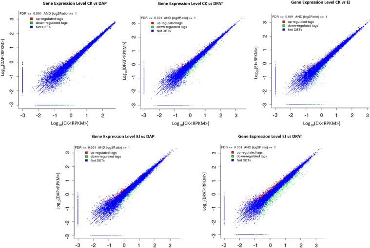
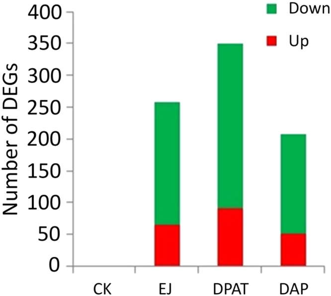
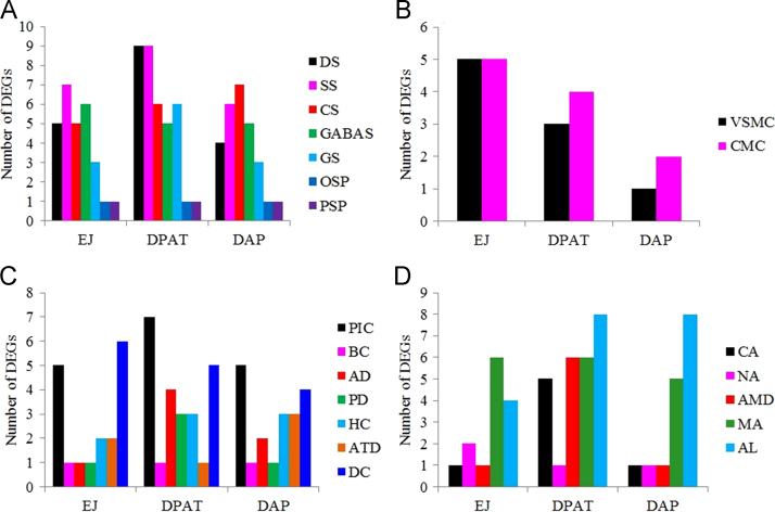
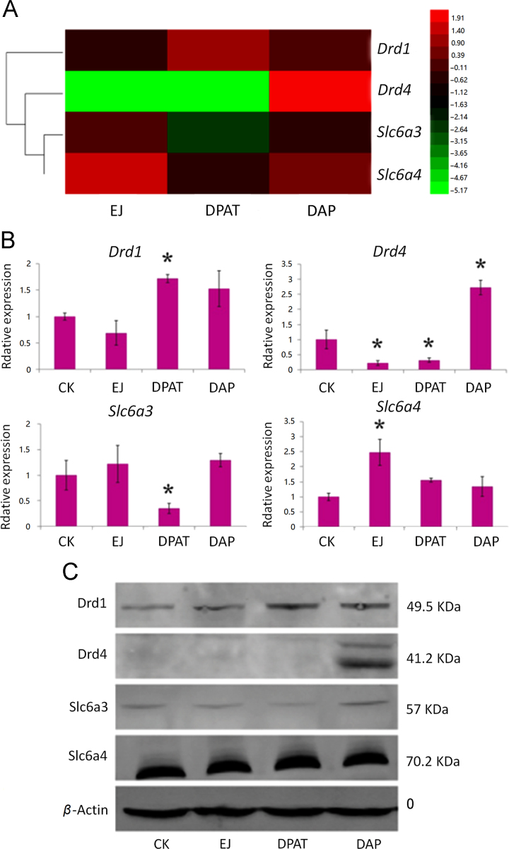
The 5-HT1A receptor agonist 8-hydroxy-2-[di-n-propylamino] tetralin (8-OH-DPAT) promotes ejaculation of male rats, whereas dapoxetine delays this process. However, the gene expression profile of the brain at ejaculation following administrationof these two compounds has not been fully elucidated. In the present study, a transcriptomic BodyMap was generated by conducting mRNA-Seq on brain samples of male Sprague-Dawley rats. The study included four groups: pre-copulatory control (CK) group, ejaculation (EJ) group, 0.5 mg/kg 8-OH-DPAT-ejaculation group (DPAT), and 60 mg/kg dapoxetine-ejaculation (DAP) group. The resulting analysis generated an average of approximately 47 million sequence reads. Significant differences in the gene expression profiles of the aforementioned groups were observed in the EJ (257 genes), DPAT (349 genes) and the DAP (207 genes) compared with the control rats. The results indicate that the expression of Drd1 and Slc6a3 was significantly different after treatment with 8-OH-DPAT, whereas the expression of Drd4 was significantly different after treatment with dapoxetine. Other genes, such as Wnt9b, Cdkn1a and Fosb, exhibited significant differences in expression after the two treatments and are related to bladder cancer, renal cell carcinoma and sexual addiction. The present study reveals the basic pattern of gene expression that was activated at ejaculation in the presence of 8-OH-DPAT or dapoxetine, providing preliminary gene expression information during rat ejaculation.

Keywords: 8-OH-DPAT; Brain; Dapoxetine; Ejaculation; Gene expression; Male rats.


Figures

fx1
Graphical abstract
Fig. 1
Figure 1
Differences in rat transcriptomic profiles. The results are depicted as scatter plots and indicate the gene expression values that were expressed as log2 (RPKM), for CK (x axis) and the other groups of rats and the concentration of compounds administered in a log scale (y axis). The plots indicate the gene expression profiles between the CK and the other groups of rats in the three different treated ejaculation stages. Red: up-regulated; Green: down-regulated. The mean value of gene expression exhibits a significant difference (FDR ≤ 0.001, fold change ≥ 2), whereas the blue color corresponds to a non-significant difference. CK, control; EJ, ejaculation; DPAT, 0.5 mg/kg 8-OH-DPT-ejaculation; DAP, 60 mg/kg dapoxetine-ejaculation.
Fig. 2
Figure 2
Percentages of differentially expressed genes (DEGs). DEG values were transformed according to the following thresholds: |log2Ratio| ≥1 and FDR ≤ 0.001. A 2-fold change or higher was considered as an up-regulation. CK, control; EJ, ejaculation; DPAT, 0.5 mg/kg 8-OH-DPT-ejaculation; DAP, 60 mg/kg dapoxetine-ejaculation.
Fig. 3
Figure 3
Histogram of gene ontology classification. The results are summarized in three main categories: cellular component, molecular function and biological process. The right y-axis indicates the number of genes in a category. The left y-axis indicates the percentage of a specific category of genes in the main category. CK, control; EJ, ejaculation; DPAT, 0.5 mg/kg 8-OH-DPT-ejaculation; DAP, 60 mg/kg dapoxetine-ejaculation.
Fig. 4
Figure 4
The changes in the number of the DEGs associated with various signaling pathways during ejaculation induced by different treatments. (A) A total of seven pathways related to the transmitter synapse and the endocrine hormone signaling pathways were notably enriched. (B) Two pathways related to muscle contraction were notably enriched. (C) Seven pathways related to the pathways in cancer, bladder cancer, Alzheimer׳s disease, Parkinson׳s disease, Hepaitis C, autoimmune thyroid and dilated cardiomyopathy were notably enriched. (D) Five pathways related to addiction were notably enriched. DS, dopaminergic synapse; SS, serotonergic synapse; CS, cholinergic synapse; GABAS, GABAergic synapse; GS, glutamatergic synapse; OSP, oxytocin signaling pathway; PSP, prolactin signaling pathway; VSUC, vascular smooth muscle contraction; CMC, cardiac muscle contraction; PIC, pathways in cancer; BC, Bladder cancer; AD, Alzheimer׳s disease; PD, Parkinson׳s disease; HC, Hepaitis C; ATD, autoimmune thyroid disease; DC, dilated cardiomyopathy; CA, Cocaine addiction; NA, Nicotine addiction; AMD, amphetamine addiction; MA, morphine addiction; AL, alcoholism. CK, control; EJ, ejaculation; DPAT, 0.5 mg/kg 8-OH-DPT; DAP, 60 mg/kg dapoxetine.
Fig. 5
Figure 5
Changes of Drd1, Drd4, Slc6a3 and Slc6a4 expression. (A) Clustering analysis of four genes measured in RNA-Seq. In RNA-Seq, expression was calculated with the log scale of RPKM values. The significant difference between CK and other groups was justified by |log2Ratio| ≥ 1, p < 0.05. (B) Relative expression of four genes measured in RT-qPCR. Gene expression was calculated using the 2−ΔΔC algorithm with CK as the control (*P < 0.05). (C) Four genes corresponding to protein measured in Western blot. The resulting four protein bands were detected clearly. CK, control; EJ, ejaculation; DPAT, 0.5 mg/kg 8-OH-DPT; DAP, 60 mg/kg dapoxetine.

Similar articles

Cited by

References

    1. Newman H.F., Reiss H., Northup J.D. Physical basis of emission, ejaculation, and orgasm in the male. Urology. 1982;19:341–350. - PubMed
    1. deGroat W.C., Booth A.M. Physiology of male sexual function. Ann Intern Med. 1980;92:329–331. - PubMed
    1. Bole-Feysot C., Goffin V., Edery M., Binart N., Kelly P.A. Prolactin (PRL) and its receptor: actions, signal transduction pathways and phenotypes observed in PRL receptor knockout mice. Endocr Rev. 1998;19:225–268. - PubMed
    1. Krüger T.H., Hartmann U., Schedlowski M. Prolactinergic and dopaminergic mechanisms underlying sexual arousal and orgasm in humans. World J Urol. 2005;23:130–138. - PubMed
    1. Hull E.M., Dominguez J.M. Getting his act together: roles of glutamate, nitric oxide, and dopamine in the medial preoptic area. Brain Res. 2006;1126:66–75. - PubMed