

Role of different imaging modalities of vascular calcification in predicting outcomes in chronic kidney disease
- PMID: 28540199
- PMCID: PMC5424431
- DOI: 10.5527/wjn.v6.i3.100
Role of different imaging modalities of vascular calcification in predicting outcomes in chronic kidney disease
Abstract
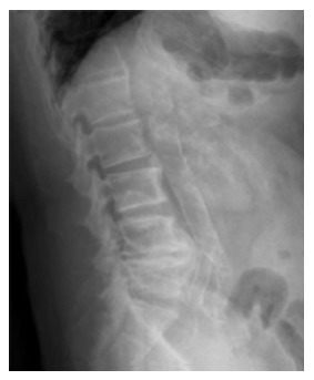
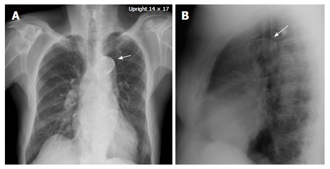
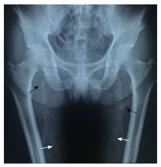
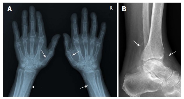
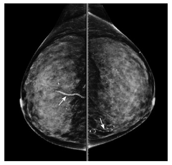
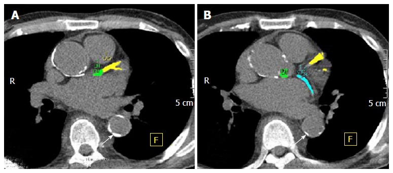
Vascular calcification (VC) is common among patients with chronic kidney disease (CKD). The severity of VC is associated with increased risk of cardiovascular events and mortality. Risk factors for VC include traditional cardiovascular risk factors as well as CKD-related risk factors such as increased calcium and phosphate load. VC is observed in arteries of all sizes from small arterioles to aorta, both in the intima and the media of arterial wall. Several imaging techniques have been utilized in the evaluation of the extent and the severity of VC. Plain radiographs are simple and readily available but with the limitation of decreased sensitivity and subjective and semi-quantitative quantification methods. Mammography, especially useful among women, offers a unique way to study breast arterial calcification, which is largely a medial-type calcification. Ultrasonography is suitable for calcification in superficial arteries. Analyses of wall thickness and lumen size are also possible. Computed tomography (CT) scan, the gold standard, is the most sensitive technique for evaluation of VC. CT scan of coronary artery calcification is not only useful for cardiovascular risk stratification but also offers an accurate and an objective analysis of the severity and progression.
Keywords: Aortic calcification; Coronary calcification; Dialysis; Hemodialysis; Mammogram; Plain X-ray; Ultrasound.
Conflict of interest statement
Conflict-of-interest statement: Authors declare no conflict of interests for this article.
Figures
References
-
- Hyman JB, Epstein FH. A study of the correlation between roentgenographic and post-mortem calcification of the aorta. Am Heart J. 1954;48:540–543. - PubMed
-
- Stamler J. The epidemiology of atherosclerotic coronary heart disease. I. Postgrad Med. 1959;25:610–622. - PubMed
-
- Witteman JC, Kok FJ, van Saase JL, Valkenburg HA. Aortic calcification as a predictor of cardiovascular mortality. Lancet. 1986;2:1120–1122. - PubMed
-
- Kauppila LI, Polak JF, Cupples LA, Hannan MT, Kiel DP, Wilson PW. New indices to classify location, severity and progression of calcific lesions in the abdominal aorta: a 25-year follow-up study. Atherosclerosis. 1997;132:245–250. - PubMed
Publication types
LinkOut - more resources
Full Text Sources
Other Literature Sources
