. 2017:2017:6895730.
doi: 10.1155/2017/6895730. Epub 2017 Apr 30.

Extracellular Signal-Regulated Kinase 5 is Required for Low-Concentration H2O2-Induced Angiogenesis of Human Umbilical Vein Endothelial Cells

Affiliations

Extracellular Signal-Regulated Kinase 5 is Required for Low-Concentration H2O2-Induced Angiogenesis of Human Umbilical Vein Endothelial Cells

Shan Jiang et al. Biomed Res Int. 2017.

Abstract

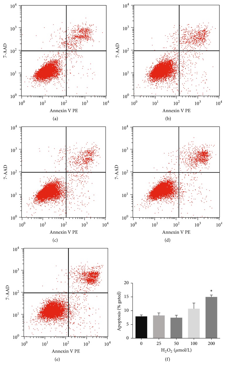
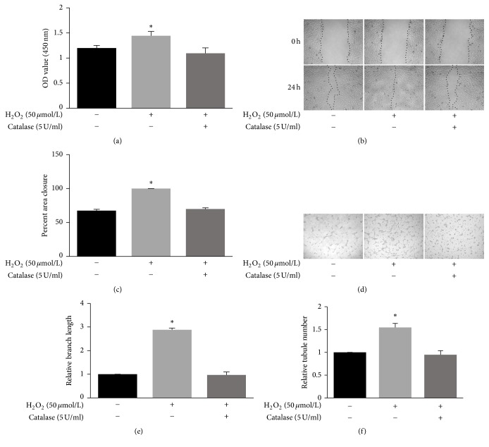
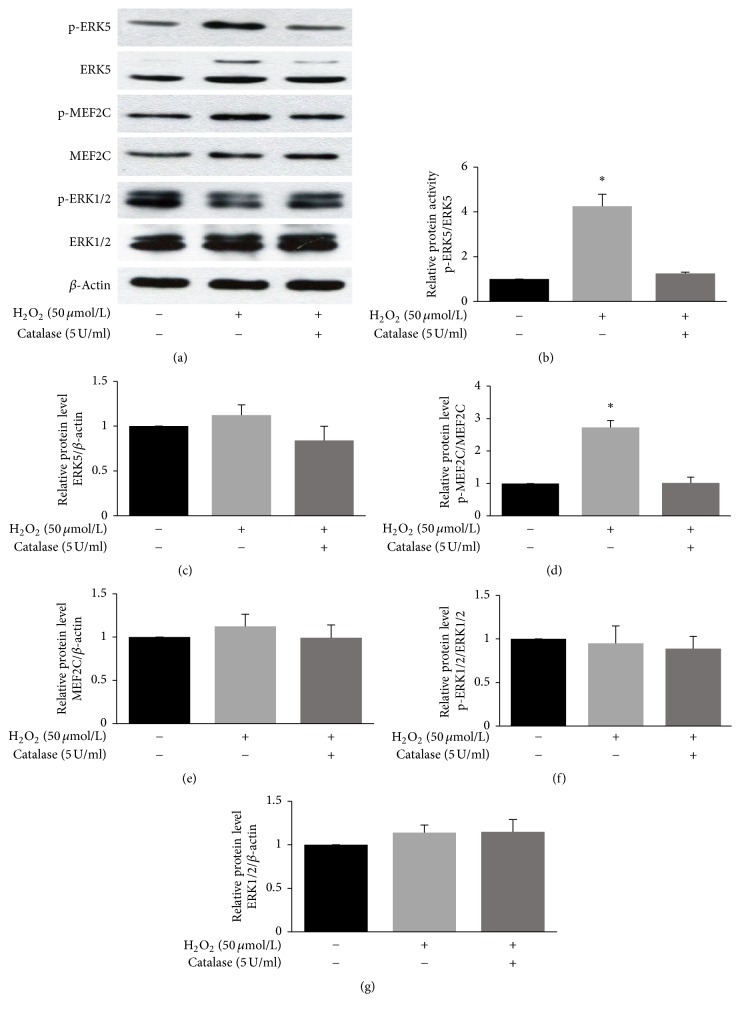
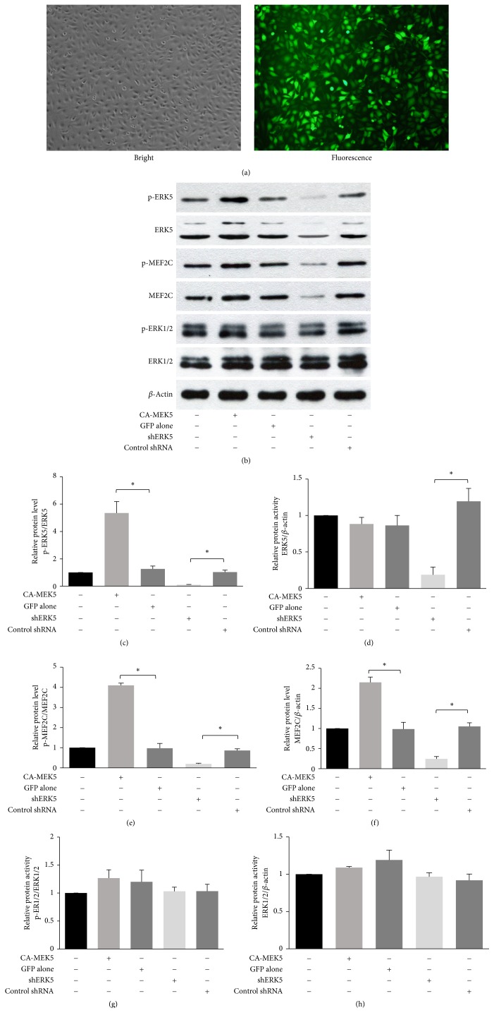
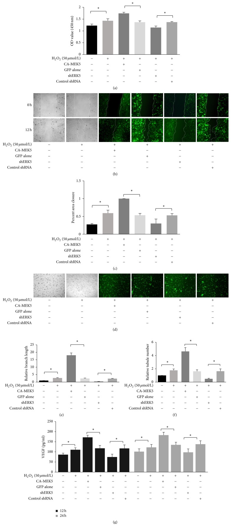
Background. The aim of this study was to assess the effects of low concentrations of H2O2 on angiogenesis of human umbilical vein endothelial cells (HUVECs) in vitro and explore the underlying mechanisms. Methods. HUVECs were cultured and stimulated with different concentrations of H2O2. Flow cytometric analysis was used to select an optimal concentration of H2O2 for the following experiments. Cell proliferation, migration, and tubule formation were evaluated by Cell Counting Kit-8 (CCK-8) assays, scratch wound assays, and Matrigel tubule formation assays, respectively. For gain and loss of function studies, constitutively active MEK5 (CA-MEK5) and ERK5 shRNA lentiviruses were used to activate or knock down extracellular signal-regulated kinase 5 (ERK5). Results. We found that low concentrations of H2O2 promoted HUVECs proliferation, migration, and tubule formation. ERK5 in HUVECs was significantly activated by H2O2. Enhanced ERK5 activity significantly amplified the proangiogenic effects of H2O2; in contrast, ERK5 knock-down abrogated the effects of H2O2. Conclusions. Our results confirmed that low concentrations of H2O2 promoted HUVECs angiogenesis in vitro, and ERK5 is an essential mediator of this process. Therefore, ERK5 may be a potential therapeutic target for promoting angiogenesis and improving graft survival.


Figures

Figure 1
Figure 1
Effects of different concentrations of H2O2 on HUVEC viability. (a–e) Scatter plots of flow cytometric results with annexin V (horizontal axis) and 7-AAD (vertical axis). Cells were labeled with annexin V and 7-AAD after a 30 min incubation with different concentrations of H2O2. The conditions were as follows: (a) 0 μmol/L H2O2 (control), (b) 25 μmol/L H2O2, (c) 50 μmol/L H2O2, (d) 100 μmol/L H2O2, and (e) 200 μmol/L H2O2. Early apoptotic cells were defined as annexin V positive/7-AAD negative. (f) Quantitative analysis of cell apoptosis with different concentrations of H2O2 (mean ± SD of six separate experiments, p < 0.05 versus control, and one-way ANOVA followed by SNK post hoc test).
Figure 2
Figure 2
Low concentrations of H2O2 promote HUVEC proliferation, migration, and tubule formation. (a) Cell proliferation assessed by CCK-8 assays. HUVECs were treated with PBS, H2O2 (50 μmol/L), or catalase (5 U/ml) and H2O2 (50 μmol/L) for 30 min. Then, 10 μl CCK-8 was added. The OD at 450 nm was measured 2.5 h later (mean ± SD of six separate experiments, p < 0.05 versus control, and one-way ANOVA followed by SNK post hoc test). (b, c) Cell migration assessed by scratch wound assays. HUVECs were seeded at confluence and cultured overnight. Following treatment with PBS, H2O2 (50 μmol/L), or catalase (5 U/ml) and H2O2 (50 μmol/L) for 30 min, a scratch was made. The wounded HUVECs monolayers were then incubated with medium containing 2% FBS for 24 h. Photographs were taken at 0 h and 24 h at fixed locations along the scratch, and the closure of the wound area was quantified (mean ± SD of six separate experiments, p < 0.05 versus control, and one-way ANOVA followed by SNK post hoc test). (d, e, f) In vitro tubule formation. Tubule formation was assayed on Matrigel. Cells were pretreated with PBS, H2O2 (50 μmol/L), or catalase (5 U/ml) and H2O2 (50 μmol/L) for 30 min and then seeded onto Matrigel-coated wells in normal medium and cultured for 4 to 8 h. Tubule formation was analyzed by measuring branch length and counting tubule number (mean ± SD of six separate experiments, p < 0.05 versus control, and one-way ANOVA followed by SNK post hoc test).
Figure 3
Figure 3
Low-concentration H2O2 induced ERK5 activation in HUVECs. (a) ERK5, p-ERK5, MEF2C, p-MEF2C, ERK1/2, and p-ERK1/2 protein levels were measured by western blotting. Following treatment with PBS, H2O2 (50 μmol/L), or catalase (5 U/ml) and H2O2 (50 μmol/L) for 30 min, cells were lysed, and the lysates were resolved by 10% or 6% SDS-PAGE. (b, c, d, e, f, g) Densitometric quantification of ERK5, p-ERK5, MEF2C, p-MEF2C, ERK1/2, and p-ERK1/2 expression (mean ± SD of three separate experiments, p < 0.05 versus control, and one-way ANOVA followed by SNK post hoc test).
Figure 4
Figure 4
Construction of high or low p-ERK5 HUVEC populations. (a) Images of lentivirus-transfected HUVECs. (b) ERK5, p-ERK5, MEF2C, p-MEF2C, ERK1/2, and p-ERK1/2 protein levels were determined by western blotting. Different cell populations were lysed, and the lysates were resolved by 10% or 6% SDS-PAGE. (c, d, e, f, g, h) Densitometric quantification of ERK5, p-ERK5, MEF2C, p-MEF2C, ERK1/2, and p-ERK1/2 expression (mean ± SD of three separate experiments, p < 0.05, and one-way ANOVA followed by SNK post hoc test).
Figure 5
Figure 5
Activated ERK5 is essential for low-concentration H2O2-induced HUVEC angiogenesis. (a) Cell proliferation assessed by CCK-8 assays. Cells were treated with PBS or H2O2 (50 μmol/L) for 30 min. Then, 10 μl CCK-8 was added. The OD value at 450 nm was measured 2.5 h later (mean ± SD of six separate experiments, p < 0.05, and one-way ANOVA followed by SNK post hoc test). (b, c) Cell migration assessed by scratch wound assays. Cells were seeded at confluence and cultured overnight. Following treatment with PBS or H2O2 (50 μmol/L) for 30 min, a scratch was made. The wounded cell monolayers were then incubated with medium containing 2% FBS for 12 h. Photographs were taken at 0 h and 12 h at fixed locations along the scratch, and closure of the wound was quantified (mean ± SD of six separate experiments, p < 0.05, and one-way ANOVA followed by SNK post hoc test). (d, e, f) In vitro tubule formation. Tubule formation was assayed on Matrigel. Cells were pretreated with PBS or H2O2 (50 μmol/L) for 30 min and then seeded onto Matrigel-coated wells in normal medium and cultured for 4 to 8 h. Tubule formation was analyzed by measuring branch length and counting tubule number (mean ± SD of six separate experiments, p < 0.05, and one-way ANOVA followed by SNK post hoc test). (g) VEGF secretion assessed by ELISA. Cells were treated with PBS or H2O2 (50 μmol/L) for 30 min; then the medium was changed to fresh normal medium. After 12 h or 24 h culture, the cell supernatant was collected, and the concentration of VEGF was determined by a human VEGF ELISA kit (mean ± SD of six separate experiments, p < 0.05, and one-way ANOVA followed by SNK post hoc test).

Similar articles

Cited by

References

    1. Kurzyk A. Angiogenesis—possibilities, problems and perspectives. Postepy Biochemii. 2015;61(1):25–34. - PubMed
    1. Panieri E., Santoro M. M. ROS signaling and redox biology in endothelial cells. Cellular and Molecular Life Sciences. 2015;72(17):3281–3303. doi: 10.1007/s00018-015-1928-9. - DOI - PMC - PubMed
    1. Oshikawa J., Kim S.-J., Furuta E., et al. Novel role of p66shc in ROS-dependent VEGF signaling and angiogenesis in endothelial cells. American Journal of Physiology—Heart and Circulatory Physiology. 2012;302(3):H724–H732. doi: 10.1152/ajpheart.00739.2011. - DOI - PMC - PubMed
    1. García-Quintans N., Sánchez-Ramos C., Tierrez A., et al. Control of endothelial function and angiogenesis by PGC-1α relies on ROS control of vascular stability. Free Radical Biology and Medicine. 2014;75(1):p. S5. doi: 10.1016/j.freeradbiomed.2014.10.836. - DOI - PubMed
    1. Kim G. D., Oh J., Park H.-J., Bae K., Lee S. K. Magnolol inhibits angiogenesis by regulating ROS-mediated apoptosis and the PI3K/AKT/mTOR signaling pathway in mES/EB-derived endothelial-like cells. International Journal of Oncology. 2013;43(2):600–610. doi: 10.3892/ijo.2013.1959. - DOI - PubMed

MeSH terms

LinkOut - more resources