. 2017 Dec;63(6):1053-1072.
doi: 10.1007/s00294-017-0702-y. Epub 2017 May 24.

Yeast ENV9 encodes a conserved lipid droplet (LD) short-chain dehydrogenase involved in LD morphology

Affiliations

Yeast ENV9 encodes a conserved lipid droplet (LD) short-chain dehydrogenase involved in LD morphology

Ikha M Siddiqah et al. Curr Genet. 2017 Dec.

Abstract

Lipid droplets (LDs) have emerged as dynamic and interactive organelles with important roles in lipid metabolism and membrane biogenesis. Here, we report that Saccharomyces cerevisiae Env9 is a novel conserved oxidoreductase involved in LD morphology. Microscopic and biochemical studies confirm localization of tagged Env9 to LDs and implicate its C-terminal hydrophobic domain (aa241-265) in its membrane association and stability. Confocal studies reveal a role for Env9 in LD morphology. Env9 positively affects both formation of large LDs upon overexpression and LD proliferation under poor carbon source. In silico bioinformatic and modeling approaches establish that ENV9 is a widely conserved member of the short-chain dehydrogenase (SDR) superfamily. Bayesian phylogenetic studies strongly support ENV9 as an ortholog of human SDR retinol dehydrogenase 12 (RDH12). Dehydrogenase activity of Env9 was confirmed by in vitro oxidoreductase assays. RDH12 mutations have been linked to Leber Congenital Amaurosis. Similar site-directed point mutations in the predicted Env9 oxidoreductase active site (N146L) or cofactor-binding site (G23-24A) abolished its reductase activity in vitro, consistent with those reported in other retinol dehydrogenases. The same residues were essential for affecting LD size and number in vivo. Taken together, our results implicate oxidoreductase activity of Env9 in its cellular role in LD morphology.

Keywords: ENV9; LD morphology; LD number; LD size; RDH12 ortholog; Short-chain dehydrogenase.


Figures
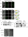
Fig. 1
Fig. 1
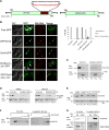
Env9 localizes to lipid droplets. a Chromosomal ENV9-GFP is expressed from its endogenous promoter and localizes to LDs in rich and minimal media. b GFP-tagged Env9 is expressed from an episomal plasmid and localizes to LDs. env9Δ cells expressing GFP-ENV9 (GFP-tag at N-terminal), ENV9-GFP (GFP-tag at C-terminal), or free GFP were induced with galactose for 5 h to express the GFP-tagged proteins, stained with Nile Red and viewed by confocal microscopy. Schematic designs of expression vectors are shown to the right. c HA-tagged Env9 localizes to LDs in subcellular fractionation experiments. Left panels env9Δ cells harboring either control plasmid or ENV9-HA plasmid were spheroplasted and subjected to differential centrifugation to obtain indicated fractions, which were analyzed by western blotting using anti-HA antibody (upper panel). Blots were stripped and reprobed with anti-ALP antibody (lower panel). Asterisk commonly reported cytoplasmic degradation product of ALP. Right panels env9Δ cells expressing ENV9-HA with or without Erg6-dsRed expressing plasmid were spheroplasted, lysed with homogenization in presence of 0.4 μg DEAE-dextran and subjected to Ficoll gradient centrifugation (1st purification). The top layer (0% Ficoll fraction) was collected and further purified using 8% Ficoll gradient (2nd purification). Fractions were collected and analyzed by western blotting using anti-HA antibody. Blots were stripped and reprobed with anti-CPY antibody (Santa Cruz, California) for detection of CPY as vacuolar marker. Same experimental samples were also subjected to separate SDS-PAGE and western blotting analysis using anti-dsRed antibody (Santa Cruz, California) for detection of Erg6 as LD marker. In addition to Erg6-dsRed, a non-specific higher molecular weight band was detected in anti-dsRed blot. d ENV9-HA is functional and complements caffeine sensitivity of env9Δ cells. env9Δ or WT cells expressing control plasmid or ENV9-HA were plated in serial dilutions on media with or without caffeine
Fig. 2
Fig. 2
A C-terminal membrane association domain anchors Env9 to lipid droplet membranes. a Schematic diagram of WT Env9 and Env9Δ241–265 deleted in the putative membrane association domain (red). b GFP-tagged Env9Δ241–265 mislocalizes to the cytoplasm and non-LD punctates. Left panels env9Δ cells, transformed with indicated plasmids, were grown in SM-Ura media to OD600 = 0.8–1.0, induced with galactose for 5 h to express GFP or GFP fusion proteins, and viewed by confocal microscopy. Right panels Quantification of data presented in left panels. *P < 0.05 compared to WT Env9-GFP. c Env9Δ241–265-HA distribution in membrane and cytosolic fractions. Cells were spheroplasted, lysed with DEAE-Dextran, and subjected to differential centrifugation to obtain indicated fractions. Fractions were collected and analyzed by western blotting using anti-HA (top panel) and anti-ALP (bottom panel, as fractionation marker) antibodies. d Env9Δ241–265-HA is a substrate for proteasomal degradation. Cells were treated with DMSO or proteosomal inhibitor MG-132 (50 μg/ml) and lysed. Fractions were analyzed by Western blotting using anti-HA and anti-ALP antibodies as described in c. e Solubilization of Env9Δ241–265-HA from membranes. Cells were treated with DMSO or MG-132 (50 μg/ml) and lysed. Soluble (S13) and membrane (P13) fractions were resuspended in the indicated solutions (TX-100—Triton X100, pH 11.5–0.1 M NaCO3, pH 11.5) and analyzed by western blotting using anti-HA antibody. f Mislocalization of Env9Δ241–265-HA in proteasome-inhibited cells. Cells were treated with MG-132 (50 μg/ml), spheroplasted, lysed with homogenization in the presence of 0.4 μg DEAE-dextran and subjected to Ficoll gradient centrifugation. Fractions were analyzed by western blotting using anti-HA antibody. g Stabilized and mislocalized Env9Δ241–265-HA is not associated with LD membranes. Cells were subjected to similar treatments as in f followed by sequential Ficoll gradient centrifugation as described in Fig. 1c. Fractions were analyzed by western blotting using anti-HA and anti-dsRed antibodies. In addition to Erg6-dsRed, a non-specific higher molecular weight band was detected in anti-dsRed blot. h Mislocalization of GFP-tagged Env9 in get1Δ and get3Δ. get1Δ or get3Δ cells, transformed with ENV9-GFP plasmid, were grown in SM-Ura media to OD600 = 0.8–1.0, induced with galactose for 5 h to express GFP fusion proteins, and viewed by confocal microscopy. i Quantification of data presented in h. *P < 0.0001 compared to WT
Fig. 2
Fig. 2
A C-terminal membrane association domain anchors Env9 to lipid droplet membranes. a Schematic diagram of WT Env9 and Env9Δ241–265 deleted in the putative membrane association domain (red). b GFP-tagged Env9Δ241–265 mislocalizes to the cytoplasm and non-LD punctates. Left panels env9Δ cells, transformed with indicated plasmids, were grown in SM-Ura media to OD600 = 0.8–1.0, induced with galactose for 5 h to express GFP or GFP fusion proteins, and viewed by confocal microscopy. Right panels Quantification of data presented in left panels. *P < 0.05 compared to WT Env9-GFP. c Env9Δ241–265-HA distribution in membrane and cytosolic fractions. Cells were spheroplasted, lysed with DEAE-Dextran, and subjected to differential centrifugation to obtain indicated fractions. Fractions were collected and analyzed by western blotting using anti-HA (top panel) and anti-ALP (bottom panel, as fractionation marker) antibodies. d Env9Δ241–265-HA is a substrate for proteasomal degradation. Cells were treated with DMSO or proteosomal inhibitor MG-132 (50 μg/ml) and lysed. Fractions were analyzed by Western blotting using anti-HA and anti-ALP antibodies as described in c. e Solubilization of Env9Δ241–265-HA from membranes. Cells were treated with DMSO or MG-132 (50 μg/ml) and lysed. Soluble (S13) and membrane (P13) fractions were resuspended in the indicated solutions (TX-100—Triton X100, pH 11.5–0.1 M NaCO3, pH 11.5) and analyzed by western blotting using anti-HA antibody. f Mislocalization of Env9Δ241–265-HA in proteasome-inhibited cells. Cells were treated with MG-132 (50 μg/ml), spheroplasted, lysed with homogenization in the presence of 0.4 μg DEAE-dextran and subjected to Ficoll gradient centrifugation. Fractions were analyzed by western blotting using anti-HA antibody. g Stabilized and mislocalized Env9Δ241–265-HA is not associated with LD membranes. Cells were subjected to similar treatments as in f followed by sequential Ficoll gradient centrifugation as described in Fig. 1c. Fractions were analyzed by western blotting using anti-HA and anti-dsRed antibodies. In addition to Erg6-dsRed, a non-specific higher molecular weight band was detected in anti-dsRed blot. h Mislocalization of GFP-tagged Env9 in get1Δ and get3Δ. get1Δ or get3Δ cells, transformed with ENV9-GFP plasmid, were grown in SM-Ura media to OD600 = 0.8–1.0, induced with galactose for 5 h to express GFP fusion proteins, and viewed by confocal microscopy. i Quantification of data presented in h. *P < 0.0001 compared to WT
Fig. 3
Fig. 3
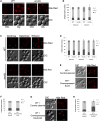
Env9 is involved in LD morphology. a ENV9 promotes glycerol-induced LD proliferation. WT and env9Δ cells were grown until stationary phase in media containing 2% dextrose or 3% glycerol, stained with Nile Red, and viewed by confocal microscopy for lipid droplet morphology. b Quantification of data presented in panel a. Approximately, 200–300 cells were scored for number of lipid droplets and categorized into ‘few lipid droplets’ (<3), ‘normal lipid droplets’ (3–7), and ‘many lipid droplets’ (>7). *P < 0.005 compared to WT under the same conditions. c ENV9 plays role in carbon stress-induced LD proliferation. WT and env9Δ cells were grown until stationary phase in media containing 2% dextrose, 2% galactose, or 3% ethanol, stained with Nile Red, and viewed by confocal microscopy for lipid droplet morphology. d Quantification of data presented in c. *P < 0.005 compared to WT under the same conditions. e Moderate overexpression of ENV9 does not affect LD morphology. WT (expressing control plasmid) or ENV9 overexpressing cells were grown until stationary phase in SM-Ura media, stained with Nile Red, and viewed by epifluorescence microscopy. f Quantification of data presented in e. g High-level overexpression of ENV9 leads to fewer and larger LDs. WT (expressing control plasmid) or ENV9-HA overexpressing cells were grown until stationary phase in SM-Ura media, stained with Nile Red, and viewed by confocal microscopy. h Quantification of data presented in g. *P < 0.0001 compared to WT
Fig. 4
Fig. 4
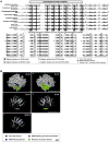
Env9 contains conserved key oxidoreductase domains. A An annotated multiple sequence alignment of Env9 amino acid sequence and representative orthologs and homologs shows well-aligned features. Eight regions either homologous or orthologous to Env9 were aligned using MAFFT alignment and visualized with geneious. The similarity of the aligned residues is indicated by a black-to-white graded scale (black corresponding to the most similarity and white to the least). Portions of the alignment containing homology-based predictions of active site and/or NADPH-binding residues are magnified below the complete multiple sequence alignment. Active site residues are indicated on Env9 by forward-slash bars underneath them, and NADPH-binding residues have black, downward-pointing triangles beneath them. B Three-dimensional representations of S. cerevisiae Env9 and H. sapiens RDH12 highlighting important structures and residues. a, b Show space-filling models of Env9, but b is rotated 180° about a longitudinal axis. c, d show ribbon models of Env9 in the same orientations as a, b, respectively. e Depicts the ribbon structure for RDH12, the human ortholog of Env9. The I-TASSER online server was used to generate a three-dimensional model in silico from the amino acid sequence for Env9. The predicted membrane-associated domain is colored green. Active site and NADPH-binding residues specific to SDRs were assigned based on multiple sequence alignment similarity or identity. Active site residues are shaded red, and NADPH-associated residues are colored blue. Two residues that are predicted to be part of both the active site and NADPH-binding region are shaded purple. N- and C-termini are labeled on the ribbon representations if visible on the model
Fig. 5
Fig. 5
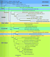
A Bayesian phylogenetic tree of Env9 and 34 of its homologs suggests its conservation in Archaea, Bacteria, and Eukaryota. The amino acid sequences of Env9 and 34 of its homologs were aligned by MAFFT multiple sequence alignment software, and this alignment was submitted to the MEGA6 program to determine an appropriate evolutionary model. The Bayesian phylogenetic tree, using H. volcanii HVO_2590 as the outgroup, was then generated from the alignment (fixed rate matrix: WAG; rate variation: invariant/gamma; gamma categories: 4; number of heated chains: 2; heated chain temperature: 0.25; chain length: 1,000,000; ASDSF: 0.05526). Sequences are shaded according to their membership to a domain or kingdom. The scale bar represents 0.4 amino acid changes. Thick branches indicate more than 90% posterior probability
Fig. 6
Fig. 6
Env9 is an oxidoreductase and requires conserved oxidoreductase domains for its enzymatic activity in vitro. Reductase activity is expressed as the rate of decrease of NADPH absorbance and expressed in percentage of the initial value. a Control reactions contained purified HMG-CoA reductase (2.5–3.5 μg), 400 μM NADPH, and 0.3 μg/μl HMG-CoA incubated for 30 min at 30 °C. P13 fractions (containing 27–30 μg protein) with or without Env9-HA were incubated with 400 μM NADPH and 0.3 μg/μl HMG-CoA (b) or 0.3 μg/μl 4-hydroxynonenal (c) for 30 min at 30 °C and rate of decrease in absorbance at 340 nm was recorded every 15 min for 30 min. d LD-enriched fractions (containing 12 μg protein) with or without Env9-HA were incubated with 400 μM NADPH and 0.3 μg/μl 4-HNE for 120 min at 30 °C. Rate of decrease in absorbance at 340 nm was recorded at 0, 30, 60, and 120 min. P13 fractions (containing 27–30 μg protein) from ENV9-overexpressing cells (ENV9-HA) or indicated mutants were assayed for reductase activities using 0.3 μg/μl HMG-CoA (e) or 0.3 μg/μl 4-hydroxynonenal (f) as described in b, c. g LD-enriched fractions (containing 12 μg protein) from ENV9-overexpressing cells (ENV9-HA) or indicated mutants were assayed for reductase activities using 0.3 μg/μl 4-HNE as described in d. Data shown are average of at least two independent experiments
Fig. 7
Fig. 7
Env9 reductase activity is required for its cellular function in LD dynamics. a Overexpression of Env9 oxidoreductase functional domain mutants did not promote LD enlargement/fusion. env9Δ cells carrying control plasmid, ENV9 overexpressing plasmid (ENV9-HA), or mutated ENV9 overexpressing plasmids (ENV9G23-24A-HA or ENV9N146L-HA), were grown to stationary phase in SM-Ura media, stained with Nile Red, and viewed by confocal microscopy for LD numbers and morphology. b Quantification of data presented in a. Data shown are representative of three independent experiments. *P < 0.0001 compared to env9Δ cells under the same conditions

References

    1. Altschul SF, Gish W, Miller W, Myers EW, Lipman DJ. Basic local alignment search tool. J Mol Biol. 1990;215:403–410. doi: 10.1016/S0022-2836(05)80360-2. - DOI - PubMed
    1. Athenstaedt K, Daum G. 1-Acyldihydroxyacetone-phosphate reductase (Ayr1p) of the yeast Saccharomyces cerevisiae encoded by the open reading frame YIL124w is a major component of lipid particles. J Biol Chem. 2000;275:235–240. doi: 10.1074/jbc.275.1.235. - DOI - PubMed
    1. Barbosa AD, Savage DB, Siniossoglou S. Lipid droplet–organelle interactions: emerging roles in lipid metabolism. Curr Opin Cell Biol. 2015;35:91–97. doi: 10.1016/j.ceb.2015.04.017. - DOI - PubMed
    1. Beeler T, Bacikova D, Gable K, Hopkins L, Johnson C, Slife H, Dunn T. The Saccharomyces cerevisiae TSC10/YBR265w gene encoding 3-ketosphinganine reductase is identified in a screen for temperature-sensitive suppressors of the Ca2+-sensitive csg2Δ mutant. J Biol Chem. 1998;273:30688–30694. - PubMed
    1. Belyaeva OV, Korkina OV, Stetsenko AV, Kim T, Nelson PS, Kedishvili NY. Biochemical properties of purified human retinol dehydrogenase 12 (RDH12): catalytic efficiency toward retinoids and C9 aldehydes and effects of cellular retinol-binding protein type I (CRBPI) and cellular retinaldehyde-binding protein (CRALBP) on the oxidation and reduction of retinoids. Biochemistry. 2005;44:7035–7047. doi: 10.1021/bi050226k. - DOI - PMC - PubMed

MeSH terms

Substances

LinkOut - more resources