. 2017 Jul;33(7):1209-1216.
doi: 10.1007/s00381-017-3466-2. Epub 2017 May 24.

Clival canal and clival foramen development in the fetal and infant basioccipital

Affiliations

Clival canal and clival foramen development in the fetal and infant basioccipital

Matthew J Zdilla. Childs Nerv Syst. 2017 Jul.

Abstract

Purpose: There is a paucity of research regarding both the development and the prevalence of the clival canal or clival foramen in fetuses. Reports that have examined child and adult populations have posited ideas for the development of the canal and foramen; however, they have done so in the absence of anatomical data from the fetal population. Therefore, the present study was performed to elucidate the development of the clival canal and foramen through the assessment of perinatal basioccipitals.

Methods: This study analyzed 104 basioccipital bones, 60 from fetuses and 44 from newborns and infants. Dorsal surfaces of basioccipitals were assessed for the presence of anatomical variation with particular attention to the presence of clival canals and foramina. Among cases in which the presence of a clival canal or clival foramen was suspected, cannulation was performed for verification.

Results: Of the 104 basioccipitals analyzed, 1 (0.96%) had a clival foramen. Clival canals were identified in seven basioccipitals (7:104; 6.73%), four of which were from fetuses. Trends in anatomical variations among basioccipitals were also identified and categorized. These categories were then evaluated relative to age in order to elucidate ontogeny. A model is presented to explain the development of the clival foramen, the clival canal, and the basioccipital, in general.

Conclusions: The presence of a clival canal or foramen should be considered even among individuals of fetal age. The findings of this osteological study suggest that the clival canal and foramen develop around vascular structures and, therefore, signify vascular connections among nearby venous plexuses.

Keywords: Anatomy; Clivus; Cranial base; Notochord.


Conflict of interest statement

Conflict of Interest: The author declares no conflict of interest.

Figures

Figure 1
Figure 1
A clival foramen and seven clival canals cannulated by thread. The clival foramen (A) opened to the ventral surface of the basioccipital. In vivo, it would represent a connection between the retropharyngeal and intracranial regions and may predispose herniation of tissues from the pharynx to the endocranium and vise-versa. Likewise, it would represent a potential path of ingress for pharyngeal infection to spread into the cranium. The clival canals where oriented transversely (B), sagittally (C,D,E), and diagonally (F,G,H).
Figure 2
Figure 2
Type A basioccipitals possessing two relatively large paramedian foveae intermediate to the lateral projections. A: 8 month-old black female fetus; B: 8 month-old black male fetus; C: 9 month-old white female fetus; D: 9 month-old white male fetus; E: newborn white female; F: newborn black male; G: newborn white female; H: 3 month-old black male infant; I: 4 month-old white female infant.
Figure 3
Figure 3
Type B basioccipitals possessing two relatively small paramedian foveolae intermediate to the lateral projections. A: 6 month-old white male fetus; B: 7 month-old black female fetus; C: 8 month-old black male fetus; D: 8 month-old black female fetus; E: 10 month-old white female fetus; F: 10 month-old white male fetus; G: newborn black male; H: newborn black female; I: 2 month-old black female infant.
Figure 4
Figure 4
Type C basioccipitals having pronounced dorsal impression that usually often has a bony strut A: 6 month-old white male fetus; B: 9 month-old black male fetus; C: 2 month-old white female infant.
Figure 5
Figure 5
Type D basioccipitals having confluent and often tortuous foveae located intermediate to the lateral projections. A: 6 month-old black male fetus; B: 8 month-old white female fetus; C: 10 month-old black female fetus; D: newborn black male; E: newborn black female; F: 3 month-old black female fetus.
Figure 6
Figure 6
Type E basioccipitals' multiple relatively large foveae are present oftentimes in addition to two paramedian foveae located intermediate to the lateral projections. A: 5 month-old white female fetus; B: 8 month-old black female fetus; C: newborn black female; D: newborn black male; E: 2 month-old black female infant; F: 2 month-old black male infant
Figure 7
Figure 7
Type F basioccipitals having one large fovea located along the midline or approximate to the midline and intermediate to the lateral projections usually accompanied by an additional fovea or foveae near the basion. A: 8 month-old black female fetus; B: 9 month-old white male fetus; C: 10 month-old black male fetus
Figure 8
Figure 8
Type G basioccipitals having a confluent fovea located intermediate to the lateral projections, roughly oriented along the coronal plane. A: 9 month-old white male fetus; B: 10 month-old black female fetus; C: 1 month-old white male infant
Figure 9
Figure 9
Type H basioccipitals having multiple foveolae located on or around the midline. One or more foveolae are located intermediate to the lateral projections. A: 8 month-old black male fetus; B: 10 month-old black male fetus; C: newborn black male
Figure 10
Figure 10
Type I basioccipitals having two roughly bilaterally symmetrical foveolae are located intermediate to the lateral projections in roughly the same coronal plane. Oftentimes, other foveolae are located intermediate to the two bilateral paramedian foveolae. A: 8 month-old white female fetus; B: 2 month-old black female infant; C: 3 month-old black male infant.
Figure 11
Figure 11
Type J basioccipitals having no centralized fossa found in the other types. A: 12 month-old black female infant; B: 14 month-old black female infant; C: 15 month-old black male infant.
Figure 12
Figure 12
Type K basioccipitals having three roughly equidistant foveae located intermediate to the lateral processes of the basioccipital. A: 6 month-old black female fetus; B: 10 month-old black male fetus; C: 10 month-old black male fetus.
Figure 13
Figure 13
Prevalence of basioccipital categories from a total of 104 basioccipitals.
Figure 14
Figure 14
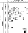
Heat map relating the relative frequency (expressed as a percentage) of basioccipital type to age grouping. To identify anatomical changes during development, relative frequencies were examined with regard to sequential age groupings. Trends were identified by assessing the most common basioccipital type in each age group and, when these types were the same or particularly similar (i.e., Type A and Type B) to the subsequent age group, they were clustered into a contiguous group outlined with thick black lines. During the 5th through 7th months of intrauterine development the basioccipital tended to have pronounced dorsal impression that usually often has a bony strut located at, or very close to, the midline forming a bifid impression which may be devoid of foveae of foveolae but usually contains shallow foveae or foveolae. Then, from the 8th month of intrauterine development to at least the 5th postnatal month, the basioccipitals tend to exhibit two paramedian foveae or foveolae intermediate to the lateral projections. Later, from the age of one year, the basioccipital resembles that of the homologous aspect of the clivus in the adult occipital bone.
Figure 15
Figure 15
Examples of variations among the foveae located at the posterior basioccipital. These fovea may occur as unilateral or bilateral rather than median structures. A: 5 month-old black female fetus with three foveae, one on the left side and two on the right side; B: 7 month-old white female fetus with multiple foveolae; C: 7 month-old white male fetus with five foveae; D: 8 month-old white female fetus with two bilateral poorly demarcated impressions; E: 8 month-old black female fetus with one fovea located on the left; F: 9 month-old white male fetus with three foveae- one median and two paramedian; G: 10 month-old white male fetus with two fovea located on the left side and one on the right side; H: newborn white male with two foveae separated by a thin bony septum; I: 3 month-old white male infant with three foveae- one approximately median and two paramedian.

Similar articles

Cited by

References

    1. Martinez CR, Hemphill JM, Hodges FJ, 3rd, Gayler BW, Nager GT, Long DM, Freeman JM. Basioccipital meningocele. AJNR Am J Neuroradiol. 1981;2:100–102. - PMC - PubMed
    1. Hemphill M, Freeman JM, Martinez CR, Nager GT, Long DM, Crumrine P. A new, treatable source of recurrent meningitis: basioccipital meningocele. Pediatrics. 1982;70:941–943. - PubMed
    1. Azizkhan RG, Cuenca RE, Powers SK. Transoral repair of a rare basioccipital meningocele in a neonate: case report. Neurosurgery. 1989;25:469–471. - PubMed
    1. Ko AL, Gabikian P, Perkins JA, Gruber DP, Avellino AM. Endoscopic repair of a rare basioccipital meningocele associated with recurrent meningitis. J Neurosurg Pediatr. 2010;6:188–192. - PubMed
    1. Mattei TA, Goulart CR. The clinical importance of basioccipital developmental defects. Childs Nerv Syst. 2012;28:501–504. - PubMed

LinkOut - more resources