Review
. 2017 Jun 13;17(12):2032-2045.
doi: 10.1039/c7lc00241f.

Finding a helix in a haystack: nucleic acid cytometry with droplet microfluidics

Affiliations
Review

Finding a helix in a haystack: nucleic acid cytometry with droplet microfluidics

Iain C Clark et al. Lab Chip. .

Abstract

Nucleic acids encode the information of life, programming cellular functions and dictating many biological outcomes. Differentiating between cells based on their nucleic acid programs is, thus, a powerful way to unravel the genetic bases of many phenotypes. This is especially important considering that most cells exist in heterogeneous populations, requiring them to be isolated before they can be studied. Existing flow cytometry techniques, however, are unable to reliably recover specific cells based on nucleic acid content. Nucleic acid cytometry is a new field built on droplet microfluidics that allows robust identification, sorting, and sequencing of cells based on specific nucleic acid biomarkers. This review highlights applications that immediately benefit from the approach, biological questions that can be addressed for the first time with it, and considerations for building successful workflows.


Figures

Figure 1
Figure 1
Nucleic acid cytometry is a general method for isolating nucleic acids based on the presence of a keyword sequence. The workflow encapsulates material, detects specific DNA or RNA sequences, and sorts droplets containing those sequences. Sorted droplets are amenable to numerous downstream analysis techniques.
Figure 2
Figure 2
Targeted comparative genomics. An example workflow: cell-free nucleic acid cytometry isolates bacterial operons based on a conserved gene sequence. The enriched material is sequenced, generating reads targeted to a specific region of interest. Bacterial operons are denoted as colored arrows, with regions of synteny highlighted in grey.
Figure 3
Figure 3
Sorting and sequencing cells based on specific RNA expression. Whole cell nucleic acid cytometry encapsulates single cells in droplets and detects RNA using reverse transcription PCR. Sorting and RNA-seq on positive drops is used to understand the transcriptional landscape of cells with disease-specific transcription, alternative splicing, or non-coding RNA.
Figure 4
Figure 4
Sorting and sequencing of extracellular vesicles. EVs are sorted based on the presence of miRNA and genome sequenced, revealing the genotype of their cellular origin.
Figure 5
Figure 5
Targeted sequencing of microbial genomes. Novel microbial clades are chosen for enrichment based on phylogenetic affiliation. Sorting and sequencing is used to understand the genomics of these uncultivable clades.
Figure 6
Figure 6
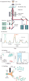
Microfluidic droplet sorting allows for the isolation drops. A) Droplet sorters use an inverted microscope to align lasers onto a microfluidic channel. B) Drops containing fluorophores are excited as they pass through the laser line. Fluorescent signals are collected in real time using photomultiplier tubes. C) Gating of positive drops is based on the intensity, shape, and width of the detected signal. Large coalesced drops produce wide signals and can be discarded. D) Sorting is achieved using a high voltage AC signal that generates a dialectrophoretic force on water droplets.

References

    1. Shipp MA, Ross KN, Tamayo P, Weng AP, Kutok JL, Aguiar RCT, Gaasenbeek M, Angelo M, Reich M, Pinkus GS, Ray TS, Koval MA, Last KW, Norton A, Lister TA, Mesirov J, Neuberg DS, Lander ES, Aster JC, Golub TR. Nat. Med. 2002;8:68–74. - PubMed
    1. Huang E, Ishida S, Pittman J, Dressman H, Bild A, Kloos M, D'Amico M, Pestell RG, West M, Nevins JR. Nat. Genet. 2003;34:226–230. - PubMed
    1. Adler AS, Lin M, Horlings H, Nuyten DSA, van de Vijver MJ, Chang HY. Nat. Genet. 2006;38:421–430. - PMC - PubMed
    1. Morissette G, Flamand L. J. Virol. 2010;84:12100–12109. - PMC - PubMed
    1. Kintses B, van Vliet LD, Devenish SR, Hollfelder F. Curr. Opin. Chem. Biol. 2010;14:548–555. - PubMed