Review
. 2017 Nov 1;29(9):401-409.
doi: 10.1093/intimm/dxx031.

Hyperferritinemia and inflammation

Affiliations
Review

Hyperferritinemia and inflammation

Kate F Kernan et al. Int Immunol. .

Abstract

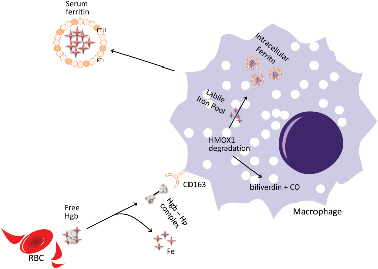
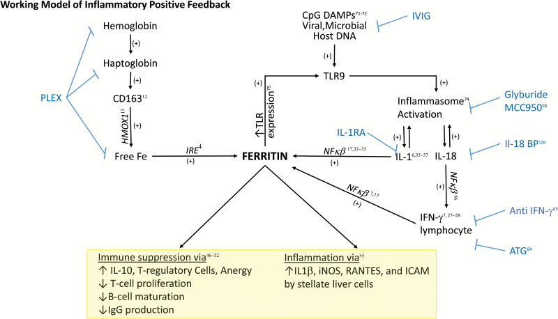
Understanding of ferritin biology has traditionally centered on its role in iron storage and homeostasis, with low ferritin levels indicative of deficiency and high levels indicative of primary or secondary hemochromatosis. However, further work has shown that iron, redox biology and inflammation are inexorably linked. During infection, increased ferritin levels represent an important host defense mechanism that deprives bacterial growth of iron and protects immune cell function. It may also be protective, limiting the production of free radicals and mediating immunomodulation. Additionally, hyperferritinemia is a key acute-phase reactants, used by clinicians as an indication for therapeutic intervention, aimed at controlling inflammation in high-risk patients. One school of thought maintains that hyperferritinemia is an 'innocent bystander' biomarker of uncontrolled inflammation that can be used to gauge effectiveness of intervention. Other schools of thought maintain that ferritin induction could be a protective negative regulatory loop. Others maintain that ferritin is a key mediator of immune dysregulation, especially in extreme hyperferritinemia, via direct immune-suppressive and pro-inflammatory effects. There is a clear need for further investigation of the role of ferritin in uncontrolled inflammatory conditions both as a biomarker and mediator of disease because its occurrence identifies patients with high mortality risk and its resolution predicts their improved survival.

Keywords: ferritin; hemophagocytic lymphohistiocytosis; hemophagocytosis; iron; macrophage activation.


Figures

Fig. 1.
Fig. 1.
The Fenton redox reaction. Ferrous Fe2+ iron is readily convertible into ferric 3+ iron, along with the production of free hydroxyl radicals and hydroxide anion. These free radicals become a source of cellular oxidative stress, damaging DNA, lipids and proteins. Ferritin molecules help sequester this free iron, preventing its participation in this reaction and subsequent free radical-mediated cellular damage.
Fig. 2.
Fig. 2.
Ferritin in RBC iron homeostasis. Macrophages phagocytize senescent RBCs directly, as well as taking up circulating hemoglobin–haptoglobin complexes (Hgb–Hp) via CD163 receptor-mediated endocytosis. This allows the macrophage to recycle iron via HMOX1, where hemoglobin is broken down into biliverdin, carbon monoxide and the free iron of the labile iron pool. It can then be incorporated into intracellular ferritin, preventing the formation of toxic free radicals. Once complexed, it is then stable for storage or transport into the plasma circulation.
Fig. 3.
Fig. 3.
Schematic of transcriptional and translational regulators of FTH expression. Ferritin expression is a highly regulated process at both the transcriptional and translational levels. (A) At the genome level, FTH ferritin gene expression has been shown to be influenced directly by transcription factors responsive to inflammatory and oxidative states including NF-κB, Nrf2 and JunD. NF-κB is thought to bind to the regulatory region FER2. P300/CBP, JunD and Nrf2 are thought to influence gene expression through interaction with the ARE, which overlaps with the FER1 promoter region (32–35). (B) The major post-transcriptional regulator of ferritin translation is iron, through its effects on the binding between IRP1 and IRP2 and the IRE in its 5′ UTR (14). Additional work has shown direct effects of IL-1β on FTH mRNA translation (36).
Fig. 4.
Fig. 4.
Proposed ferritin-mediated feed-forward inflammatory loop. In the setting of ongoing infection, CpG DNA from viral or bacterial pathogens triggers TLR9-mediated signaling. This TLR9 stimulation has been shown to activate inflammasome activity, leading to IL-1β and IL-18 production (71, 74). IL-1β activation can then in turn increase the translation of FTH mRNA through direct interaction with the 5′ UTR (36). Through ferritin’s induction of IL-1β (45) and TLR9 (75), a positive feed back loop would ensue where the generation of ferritin downstream of TLR9 activation would lead to increased amplification of inflammatory signals. Parallel to CpG DAMP-related signaling, infection would be expected to increase the production of free hemoglobin, hemoglobin–haptoglobin complexes (9, 11) and activated CD163+ macrophages, a major cellular producer of ferritin (12, 43). This increased ferritin production could then further amplify the inflammatory loop (12, 43) as well as directly alter lymphocyte function (47–53). Blue text refers to potential therapeutics targeted to steps in the pathway, including using intravenous gamma globulin (IVIG) to clear viral infection and CpG DNA, using plasma exchange (PLEX) for clearance of free hemoglobin and hemoglobin–haptoglobin complexes, as well as free ferritin, using specific anti-IFN-γ antibodies, IL-1RA or IL-18-binding protein (IL-18 BP) or using glyburide to target inflammasome activation.

Similar articles

Cited by

References

    1. Cricthon R. R. and Chaloteaux-Wauters M. 1987. Iron transport and storage. Eur. J. Biochem. 164:485. - PubMed
    1. Gozzelino R. and Soares M. P. 2014. Coupling heme and iron metabolism via ferritin H chain. Antioxid. Redox Signal. 20:1754. - PMC - PubMed
    1. Cohen L. A., Gutierrez L., Weiss A. et al. . 2010. Serum ferritin is derived primarily from macrophages through a nonclassical secretory pathway. Blood 116:1574. - PubMed
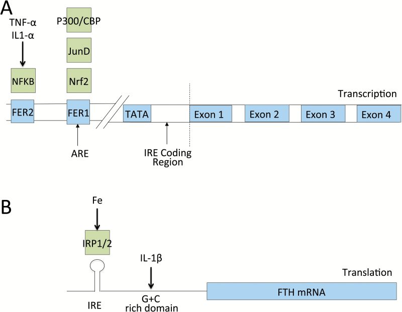
    1. Kato J., Kobune M., Ohkubo S. et al. . 2007. Iron/IRP-1-dependent regulation of mRNA expression for transferrin receptor, DMT1 and ferritin during human erythroid differentiation. Exp. Hematol. 35:879. - PubMed
    1. Harrison P. M. and Arosio P. 1996. The ferritins: molecular properties, iron storage function and cellular regulation. Biochim. Biophys. Acta 1275:161. - PubMed

Publication types

Supplementary concepts