

CORRELATION OF STRUCTURAL AND FUNCTIONAL OUTCOME MEASURES IN A PHASE ONE TRIAL OF CILIARY NEUROTROPHIC FACTOR IN TYPE 2 IDIOPATHIC MACULAR TELANGIECTASIA
- PMID: 28541963
- PMCID: PMC5700868
- DOI: 10.1097/IAE.0000000000001706
CORRELATION OF STRUCTURAL AND FUNCTIONAL OUTCOME MEASURES IN A PHASE ONE TRIAL OF CILIARY NEUROTROPHIC FACTOR IN TYPE 2 IDIOPATHIC MACULAR TELANGIECTASIA
Abstract
Purpose: Macular telangiectasia Type 2 is a bilateral, progressive, potentially blinding retinal disease characterized by both vascular and neurodegenerative signs. Both the area of the break in the ellipsoid zone seen in "en face" optical coherence tomographic (OCT) images and microperimetric focal retinal sensitivity loss have been proposed as potential measures of progression in macular telangiectasia. The authors aimed to assess the characteristics and interrelationship of these structural and functional disease markers from the data collected in a phase one clinical trial of ciliary neurotrophic factor in macular telangiectasia.
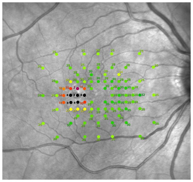
Methods: Orthogonal topographic (en face) maps of the ellipsoid zone were generated from Heidelberg Spectralis OCT volume scans (15 × 10° area, 30-μm B-scan intervals) or Zeiss Cirrus HD-OCT 4000 512 × 128 cube scans. Mesopic microperimetry was performed on CenterVue MAIA perimeters, using a Goldmann III stimulus in a custom test grid. Structural and functional data were analyzed by two methods: by calculating aggregate loss and by simple thresholding. The alignment quality of structural and functional data was also evaluated.
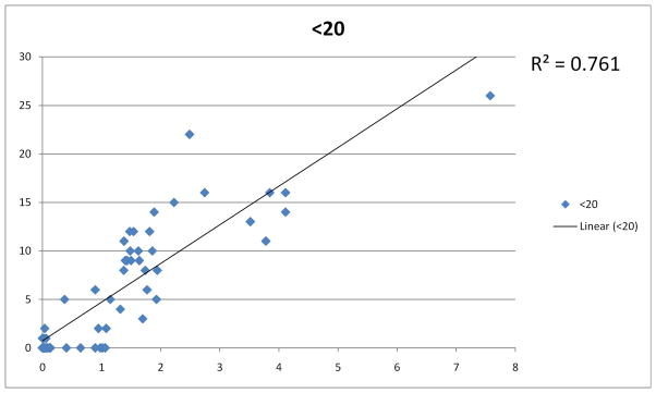
Results: Overall, the break area showed a good correlation with aggregate sensitivity loss (ρ = 0.834, P < 0.0001, 95% confidence interval 0.716-0.906) but also with the number of test points below a threshold value (e.g., <20 dB: ρ = 0.843, P < 0.0001, 95% confidence interval 0.755-0.902). Significant misalignment of the MAIA test grid was apparent in 13/48 visits of 7/14 eyes.
Conclusion: The authors found a good correlation between ellipsoid zone break area and function loss. En face OCT mapping of the ellipsoid zone appears to demonstrate structural change before mesopic microperimetry can detect a focal loss of retinal sensitivity. Thresholding offers a quick alternative to calculating aggregate sensitivity loss.
Conflict of interest statement
Figures
Similar articles
-
Correlation Between Macular Integrity Assessment and Optical Coherence Tomography Imaging of Ellipsoid Zone in Macular Telangiectasia Type 2.Invest Ophthalmol Vis Sci. 2017 May 1;58(6):BIO291-BIO299. doi: 10.1167/iovs.17-21834. Invest Ophthalmol Vis Sci. 2017. PMID: 28973315 Free PMC article. Clinical Trial.
-
LONGITUDINAL CORRELATION OF ELLIPSOID ZONE LOSS AND FUNCTIONAL LOSS IN MACULAR TELANGIECTASIA TYPE 2.Retina. 2018 Jan;38 Suppl 1(Suppl 1):S20-S26. doi: 10.1097/IAE.0000000000001715. Retina. 2018. PMID: 28541959 Free PMC article.
-
Microperimetry Features of Geographic Atrophy Identified With En Face Optical Coherence Tomography.JAMA Ophthalmol. 2016 Aug 1;134(8):873-9. doi: 10.1001/jamaophthalmol.2016.1535. JAMA Ophthalmol. 2016. PMID: 27253760
-
Functional Relevance of Hyper-Reflectivity in Macular Telangiectasia Type 2.Invest Ophthalmol Vis Sci. 2021 Mar 1;62(3):6. doi: 10.1167/iovs.62.3.6. Invest Ophthalmol Vis Sci. 2021. PMID: 33661283 Free PMC article. Clinical Trial.
-
Progression characteristics of ellipsoid zone loss in macular telangiectasia type 2.Acta Ophthalmol. 2019 Nov;97(7):e998-e1005. doi: 10.1111/aos.14110. Epub 2019 Apr 9. Acta Ophthalmol. 2019. PMID: 30968592 Free PMC article.
Cited by
-
Global Connections to Study Idiopathic Macular Telangiectasia Type 2.Retina. 2018 Jan;38 Suppl 1(Suppl 1):S3-S7. doi: 10.1097/IAE.0000000000001921. Retina. 2018. PMID: 29190235 Free PMC article. No abstract available.
-
Retinal cavitations in macular telangiectasia type 2 (MacTel): longitudinal structure-function correlations.Br J Ophthalmol. 2021 Jan;105(1):109-112. doi: 10.1136/bjophthalmol-2019-315416. Epub 2020 Mar 9. Br J Ophthalmol. 2021. PMID: 32152145 Free PMC article. Clinical Trial.
-
Estimating Retinal Sensitivity Using Optical Coherence Tomography With Deep-Learning Algorithms in Macular Telangiectasia Type 2.JAMA Netw Open. 2019 Feb 1;2(2):e188029. doi: 10.1001/jamanetworkopen.2018.8029. JAMA Netw Open. 2019. PMID: 30735236 Free PMC article.
-
Looking Ahead: Visual and Anatomical Endpoints in Future Trials of Diabetic Macular Ischemia.Ophthalmologica. 2021;244(5):451-464. doi: 10.1159/000515406. Epub 2021 Feb 24. Ophthalmologica. 2021. PMID: 33626529 Free PMC article. Review.
-
Performance of a Defect-Mapping Microperimetry Approach for Characterizing Progressive Changes in Deep Scotomas.Transl Vis Sci Technol. 2019 Aug 1;8(4):16. doi: 10.1167/tvst.8.4.16. eCollection 2019 Jul. Transl Vis Sci Technol. 2019. PMID: 31388468 Free PMC article.
References
-
- Charbel Issa P, Kupitz EH, Heeren TF, Holz FG. Treatment for Macular Telangiectasia Type 2. Developments in ophthalmology. 2016;55:189–195. - PubMed
Publication types
MeSH terms
Substances
Supplementary concepts
Grants and funding
LinkOut - more resources
Full Text Sources
Other Literature Sources
Research Materials
Miscellaneous
