. 2017 Aug;28(7):771-780.
doi: 10.1097/CAD.0000000000000516.

Hypoxia as a target for drug combination therapy of liver cancer

Affiliations

Hypoxia as a target for drug combination therapy of liver cancer

Cressida Bowyer et al. Anticancer Drugs. 2017 Aug.

Abstract

Hepatocellular carcinoma (HCC) is the third most frequent cause of cancer deaths worldwide. The standard of care for intermediate HCC is transarterial chemoembolization, which combines tumour embolization with locoregional delivery of the chemotherapeutic doxorubicin. Embolization therapies induce hypoxia, leading to the escape and proliferation of hypoxia-adapted cancer cells. The transcription factor that orchestrates responses to hypoxia is hypoxia-inducible factor 1 (HIF-1). The aim of this work is to show that targeting HIF-1 with combined drug therapy presents an opportunity for improving outcomes for HCC treatment. HepG2 cells were cultured under normoxic and hypoxic conditions exposed to doxorubicin, rapamycin and combinations thereof, and analyzed for viability and the expression of hypoxia-induced HIF-1α in response to these treatments. A pilot study was carried out to evaluate the antitumour effects of these drug combinations delivered from drug-eluting beads in vivo using an ectopic xenograft murine model of HCC. A therapeutic doxorubicin concentration that inhibits the viability of normoxic and hypoxic HepG2 cells and above which hypoxic cells are chemoresistant was identified, together with the lowest effective dose of rapamycin against normoxic and hypoxic HepG2 cells. It was shown that combinations of rapamycin and doxorubicin are more effective than doxorubicin alone. Western Blotting indicated that both doxorubicin and rapamycin inhibit hypoxia-induced accumulation of HIF-1α. Combination treatments were more effective in vivo than either treatment alone. mTOR inhibition can improve outcomes of doxorubicin treatment in HCC.


Figures
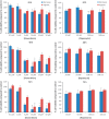
Fig. 1
Fig. 1
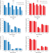
The effects of (a) doxorubicin and (b) rapamycin on the viability of HepG2 cells cultured under normoxic and hypoxic conditions. HepG2 cells were seeded onto 96-well plates (1×10⁴ cells/well) and incubated overnight. Plates were then exposed to doxorubicin or rapamycin and incubated for 24, 48 and 72 h under normoxic or hypoxic conditions. Cell viability was estimated using the MTS assay and normalized to untreated control. Data points represent mean±SEM from at least three independent experiments. For statistical analysis of doxorubicin treatments, analysis of variance with Tamhane post-hoc comparisons was used; * denotes a significant decrease in cell viability compared with the control P<0.01. For statistical analysis of rapamycin treatment, a one-tailed t-test was carried out; ** denotes a significant decrease in cell viability compared with the control P<0.05.
Fig. 2
Fig. 2
The effects of doxorubicin+10 nmol/l rapamycin on the viability of HepG2 cells cultured under (a) normoxic and (b) hypoxic conditions. HepG2 cells were seeded onto 96-well plates (1×10⁴ cells/well) and incubated overnight. Plates were then exposed to doxorubicin+10 nmol/l rapamycin and incubated for 24, 48 and 72 h under normoxic or hypoxic conditions. Cell viability was estimated using the MTS assay and normalized to untreated control. Data points represent mean±SEM from at least three independent experiments. For statistical analysis, analysis of variance was carried out. *P<0.000, **P<0.05.
Fig. 3
Fig. 3
Nuclear accumulation of hypoxia-inducible factor 1α (HIF-1α) after doxorubicin and rapamycin treatments. Normoxic and hypoxic HepG2 cells were exposed to doxorubicin or rapamycin for 24 h. Nuclear extracts were fractionated on a 10% SDS-PAGE gel, transferred to a PVDF membrane and probed with anti-HIF-1α antibodies. Proteins were visualized using chemiluminescence. The membrane was stripped and reprobed using antibodies against the nuclear house-keeping protein Lamin B1. Protein levels were quantified using densitometry analysis. HIF-1α was normalized to Lamin. Fold change compared with untreated hypoxic cells was calculated. Statistical analysis was carried out using a t-test. *P<0.05, **P<0.01.
Fig. 4
Fig. 4
Immunohistochemistry staining for hypoxia-inducible factor 1 (HIF-1) in normoxic and hypoxic HepG2 cells. Cells were seeded onto chamber slides. HepG2 cells were seeded onto chamber slides and incubated under normoxic conditions until confluence was 60%. The cells were then incubated under either (a) normoxic conditions or (b) hypoxic conditions for 24 h. The cells were incubated overnight with antibodies to HIF-1α and then incubated with the TRITC-conjugated secondary antibody. 4′,6-Diamidino-2-phenylindole staining was used to visualize the nucleus. Images are taken at ×100 magnification. HIF-1α is absent in normoxic cells and present in hypoxic cells.
Fig. 5
Fig. 5
Antitumoural activity of doxorubicin and rapamycin treatments in a mouse model of hepatocellular carcinoma. In all, 5×10⁶ HepG2 cells were subcutaneously implanted in NMRI: nu/nu mice (day 0). Tumour was palpable at day 23 after implantation and treatment was initiated. Rapamycin was administered by gavage at a dose of 1 mg/kg/day. 100 µl of beads loaded as specified was injected adjacent to the tumour. Tumour volume was measured at days 23, 25, 28, 30, 32, 35, 37, 42 and 45. Data shown represent the mean value of three replicates per group±SEM, apart from the control group, where data represent the mean of two replicates±SEM. Statistical analysis was carried out using analysis of variance analysis. *P≤0.05; **P≤0.01. DOXDEB, doxorubicin-loaded beads; RAPADEB, rapamycin-loaded beads; RAPADOXDEB, doxorubicin and rapamycin co-loaded beads.
Fig. 6
Fig. 6
Effects of doxorubicin and rapamycin treatments on tumour weight at day 45 in a mouse model of hepatocellular carcinoma. Mice were euthanized at day 45, and the tumours were excised and weighed. Data represent mean±SEM. Because of the small sample sizes, statistical analysis was not carried out. DOXDEB, doxorubicin-loaded beads; p.o., orally; RAPADEB, rapamycin-loaded beads; RAPADOXDEB, doxorubicin and rapamycin co-loaded beads.

Similar articles

Cited by

References

    1. Parkin DM, Bray F, Ferlay J, Pisani P. Global cancer statistics, 2002. CA Cancer J Clin 2005; 55:74–108. - PubMed
    1. Bruix J, Sala M, Llovet JM. Chemoembolization for hepatocellular carcinoma. Gastroenterology 2004; 127 (Suppl 1):S179–S188. - PubMed
    1. Ackerman NB. Experimental studies on the circulation dynamics of intrahepatic tumor blood supply. Cancer 1972; 29:435–439. - PubMed
    1. Breedis C, Young G. The blood supply of neoplasms in the liver. Am J Pathol 1954; 30:969–977. - PMC - PubMed
    1. Varela M, Real MI, Burrel M, Forner A, Sala M, Brunet M, et al. Chemoembolization of hepatocellular carcinoma with drug eluting beads: efficacy and doxorubicin pharmacokinetics. J Hepatol 2007; 46:474–481. - PubMed

Publication types

MeSH terms