

Structural models of the different trimers present in the core of phycobilisomes from Gracilaria chilensis based on crystal structures and sequences
- PMID: 28542288
- PMCID: PMC5436742
- DOI: 10.1371/journal.pone.0177540
Structural models of the different trimers present in the core of phycobilisomes from Gracilaria chilensis based on crystal structures and sequences
Abstract
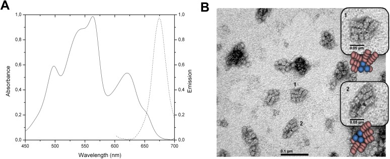
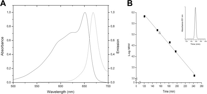
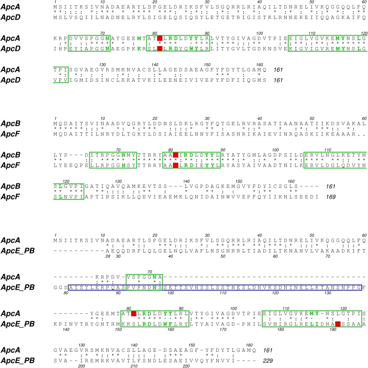
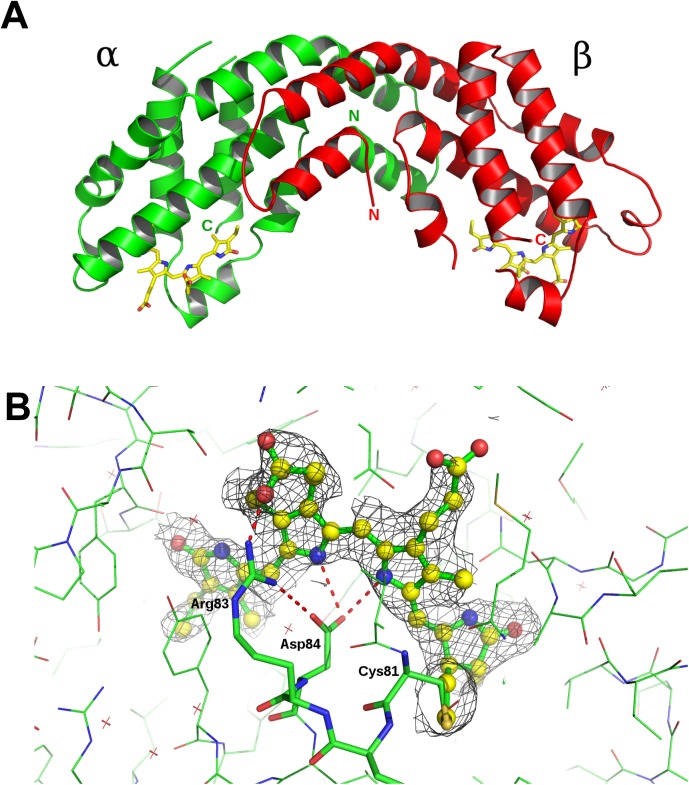
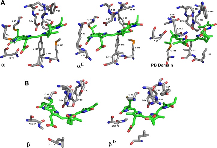
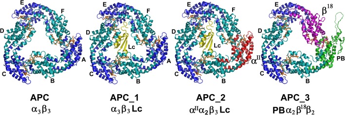
Phycobilisomes (PBS) are accessory light harvesting protein complexes that directionally transfer energy towards photosystems. Phycobilisomes are organized in a central core and rods radiating from it. Components of phycobilisomes in Gracilaria chilensis (Gch) are Phycobiliproteins (PBPs), Phycoerythrin (PE), and Phycocyanin (PC) in the rods, while Allophycocyanin (APC) is found in the core, and linker proteins (L). The function of such complexes depends on the structure of each component and their interaction. The core of PBS from cyanobacteria is mainly composed by cylinders of trimers of α and β subunits forming heterodimers of Allophycocyanin, and other components of the core including subunits αII and β18. As for the linkers, Linker core (LC) and Linker core membrane (LCM) are essential for the final emission towards photoreaction centers. Since we have previously focused our studies on the rods of the PBS, in the present article we investigated the components of the core in the phycobilisome from the eukaryotic algae, Gracilaria chilensis and their organization into trimers. Transmission electron microscopy provided the information for a three cylinders core, while the three dimensional structure of Allophycocyanin purified from Gch was determined by X-ray diffraction method and the biological unit was determined as a trimer by size exclusion chromatography. The protein sequences of all the components of the core were obtained by sequencing the corresponding genes and their expression confirmed by transcriptomic analysis. These subunits have seldom been reported in red algae, but not in Gracilaria chilensis. The subunits not present in the crystallographic structure were modeled to build the different composition of trimers. This article proposes structural models for the different types of trimers present in the core of phycobilisomes of Gch as a first step towards the final model for energy transfer in this system.
Conflict of interest statement
Figures
References
-
- Glazer AN. Light guides. Directional energy transfer in a photosynthetic antenna. J Biol Chem. 1989;264: 1–4. - PubMed
-
- MacColl R. Cyanobacterial Phycobilisomes. J Struct Biol. 1998;124: 311–334. doi: 10.1006/jsbi.1998.4062 - DOI - PubMed
-
- Adir N. Elucidation of the molecular structures of components of the phycobilisome: reconstructing a giant. Photosynth Res. 2005;85: 15–32. doi: 10.1007/s11120-004-2143-y - DOI - PubMed
-
- Sun L, Wang S, Zhao M, Fu X, Gong X, Chen M, et al. Phycobilisomes from Cyanobacteria. Handbook on Cyanobacteria: Biochemistry, Biotechnology and Applications. Nova Science Publishers; 2009. pp. 105–160.
-
- Sidler WA. Phycobilisome and Phycobiliprotein Structures In: Bryant DA, editor. The Molecular Biology of Cyanobacteria. Springer Netherlands; 1994. pp. 139–216.
MeSH terms
Substances
LinkOut - more resources
Full Text Sources
Other Literature Sources
Research Materials
