

Structural parameters associated with location of peaks of peripapillary retinal nerve fiber layer thickness in young healthy eyes
- PMID: 28542289
- PMCID: PMC5444611
- DOI: 10.1371/journal.pone.0177247
Structural parameters associated with location of peaks of peripapillary retinal nerve fiber layer thickness in young healthy eyes
Abstract
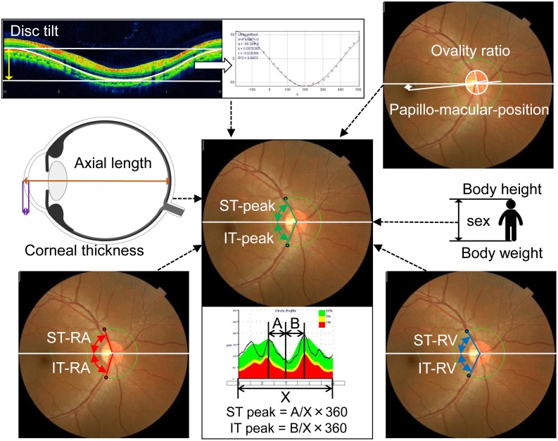
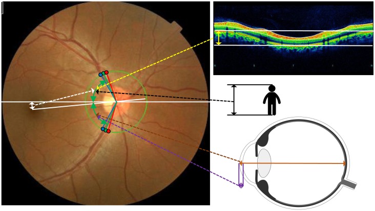
The location of the peaks of the circumpapillary retinal nerve fiber layer (cpRNFL) thickness is affected by several ocular parameters. In this study, we have generated equations that can determine the peaks of the cpRNFL. This study was a prospective, observational, cross sectional study of 118 healthy right eyes. The axial length, optic disc tilt, superiortemporal (ST)- and inferiortemporal (IT)-peaks of the cpRNFL thickness, and angles of the ST and IT retinal arteries (RA) and veins (RV) were determined. The correlations between the location of the ST- and IT-peaks and ocular structural parameters and the sex, body height and weight were calculated. The best fit equations to generate the location of the ST/IT-peaks were determined using corrected-Akaike Information Criteria. The location of the ST-peak was 0.72+(0.40 x ST-RA)+(0.27 x ST-RV)+(0.14 x height)-(0.47 x papillo-macular-position)-(0.11 x disc tilt) with a coefficient of correlation of 0.61 (P<0.0001). The location of the IT-peak was 21.88+(0.53 x IT-RA)+(0.15 x IT-RV)+(0.041 x corneal thickness)-(1.00 x axial length) with a coefficient of correlation of 0.59 (P<0.0001). The location of ST/IT peaks is determined by different parameters of the ocular structure. These equations allow clinicians to obtain an accurate location of the peaks for a more accurate diagnosis of glaucoma.
Conflict of interest statement
Figures
References
-
- Quigley HA, Katz J, Derick RJ, Gilbert D, Sommer A. An evaluation of optic disc and nerve fiber layer examinations in monitoring progression of early glaucoma damage. Ophthalmology 1992;99:19–28 - PubMed
-
- Sommer A, Katz J, Quigley HA, Miller NR, Robin AL, Richter RC, et al. Clinically detectable nerve fiber atrophy precedes the onset of glaucomatous field loss. Arch Ophthalmol 1991;109:77–83 - PubMed
-
- Blumenthal EZ, Williams JM, Weinreb RN, Girkin CA, Berry CC,Linda M, Zangwill LM. Reproducibility of nerve fiber layer thickness measurements by use of optical coherence tomography. Ophthalmology. 2000;107:2278–82. - PubMed
Publication types
MeSH terms
LinkOut - more resources
Full Text Sources
Other Literature Sources
