. 2017 May 18;12(5):e0177995.
doi: 10.1371/journal.pone.0177995. eCollection 2017.

Ghrelin modulates testicular damage in a cryptorchid mouse model

Affiliations

Ghrelin modulates testicular damage in a cryptorchid mouse model

Enrica Bianchi et al. PLoS One. .

Abstract

Cryptorchidism or undescended testis (UDT) is a common congenital abnormality associated with increased risk for developing male infertility and testicular cancer. This study elucidated the effects of endogenous ghrelin or growth hormone secretagogue receptor (GHSR) deletion on mouse reproductive performance and evaluated the ability of ghrelin to prevent testicular damage in a surgical cryptorchid mouse model. Reciprocal matings with heterozygous/homozygous ghrelin and GHSR knockout mice were performed. Litter size and germ cell apoptosis were recorded and testicular histological evaluations were performed. Wild type and GHSR knockout adult mice were subjected to creation of unilateral surgical cryptorchidism that is a model of heat-induced germ cell death. All mice were randomly separated into two groups: treatment with ghrelin or with saline. To assess testicular damage, the following endpoints were evaluated: testis weight, seminiferous tubule diameter, percentage of seminiferous tubules with spermatids and with multinucleated giant cells. Our findings indicated that endogenous ghrelin deletion altered male fertility. Moreover, ghrelin treatment ameliorated the testicular weight changes caused by surgically induced cryptorchidism. Testicular histopathology revealed a significant preservation of spermatogenesis and seminiferous tubule diameter in the ghrelin-treated cryptorchid testes of GHSR KO mice, suggesting that this protective effect of ghrelin was mediated by an unknown mechanism. In conclusion, ghrelin therapy could be useful to suppress testicular damage induced by hyperthermia, and future investigations will focus on the underlying mechanisms by which ghrelin mitigates testicular damage.


Conflict of interest statement

Competing Interests: Kim Boekelheide and Susan Hall both own stock in Semma Therapeutics (formerly CytoSolv, Inc.) a non publicly-traded small biotechnology start-up company involved in the development of a cell-based therapy for diabetes. This does not alter our adherence to PLOS ONE policies on sharing data and materials.

Figures

Fig 1
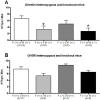
Fig 1. Evaluation of the reproductive performance of ghrelin KO and GHSR KO mice.
A) Breeding pairs of ghrelin heterozygous (+/-) and knockout (-/-) mice were co-housed for 10 days and the number of pups per litter was determined. Breeding pairs that included ghrelin KO males showed a significant decrease in the number of pups. B) Breeding pairs of GHSR heterozygous (+/-) and knockout (-/-) mice were co-housed for 10 days and the number of pups per litter was determined. No significant differences were detected among the different mating groups. Only breeding pairs with mice between 2 and 8 months old were included. Data were analyzed by one-way ANOVA multiple comparisons with Bonferroni's test and expressed as mean ± SEM (* p<0.05), and compared to the control (+/-) x (+/-) matings.
Fig 2
Fig 2. Increased testicular weight in ghrelin KO mice.
A) Testicular weights and picture of WT, GHSR KO and Ghrelin KO testes. The right testis weight was significantly increased in ghrelin KO mice compared to the WT and GHSR KO mice (80 day old). No significant differences were detected in testis weight between WT mice and GHSR KO mice. Photomicrographs of testis cross-sections from 80 day old WT mice (B), ghrelin KO mice (C) and GHSR KO mice (D). Scale bar = 200 μm. Data were analyzed by one-way ANOVA multiple comparisons Bonferroni's test and expressed as mean ± SEM (** p<0.01).
Fig 3
Fig 3. Increased germ cell apoptosis in ghrelin KO mice.
Apoptotic germ cells were detected by TUNEL analysis in wild type (A), ghrelin KO (B) and GHSR KO (C) mice. The arrows indicate apoptotic germ cells. Scale bar = 200μm. D) Quantitation of TUNEL-positive cells in wild type, ghrelin KO and GHSR KO mice testes. The data were expressed as the percentage of round seminiferous tubules (ratio major axis/minor axis < 1.5) with more than 3 TUNEL-positive cells. Ghrelin KO mice showed a significant increase in the percentage of seminiferous tubules with >3 TUNEL-positive cells compared to wild type mice. Data were analyzed by one-way ANOVA multiple comparisons Bonferroni's test and expressed as mean ± SEM (* p<0.05).
Fig 4
Fig 4. Ghrelin prevented testicular weight change in wild type and GHSR KO cryptorchid mice.
At 1 day-post surgery, testis weight was significantly increased in saline-treated cryptorchid testes compared to saline-treated normal testes. This initial testicular swelling was prevented by ghrelin treatment (A). At 4 days post-surgery, testis weight was significantly decreased in saline-treated cryptorchid testes compared to saline-treated normal testes, and this effect was ameliorated by ghrelin treatment (B). The normal testis weight was significantly increased in saline-treated GHSR KO mice compared to the saline-treated wild type mice (C). Ghrelin administration significantly increased testis weight compared to the respective controls in both wild type and GHSR KO cryptorchid mice at 20 days post-surgery (D). Data were analyzed by one-way ANOVA multiple comparisons with Fisher’s LSD test and expressed as mean ± SEM (* p<0.05, ** p<0.01, *** p<0.001, **** p<0.0001).
Fig 5
Fig 5. Histopathological analysis of ghrelin and saline-treated cryptorchid testes excised from wild type and GHSR KO mice at 20 days post-surgery.
Cryptorchid testes from saline-treated wild type and GHSR KO mice showed degeneration of seminiferous tubules with evidence of degenerating multinucleated giant cells (arrow) at 20 days post-surgery (A, C). Ghrelin-exposed animals revealed an increase in the number of spermatids (circle) in the seminiferous tubules and a reduction in the number of multinucleated giant cells compared to the control mice (B,D). Bars = 200 μM; periodic acid Schiff and hematoxylin staining.
Fig 6
Fig 6. Quantification of spermatid-containing tubules, seminiferous tubule diameter and multinucleated giant cells (MnGC) in cryptorchid testes of wild type and GHSR KO mice.
A) The number of tubules containing spermatids was significantly higher in cryptorchid testes of ghrelin-treated GHSR KO mice than in the control. B) No significant differences in the number of tubules containing MnGC were detected between ghrelin- and saline-treated cryptorchid testes. C) Ghrelin-treated GHSR KO mice showed significant increase in minor axis seminiferous tubule diameter compared to the control. Data were analyzed by one-way ANOVA multiple comparisons with Fisher’s LSD test and expressed as mean ± SEM (** p<0.01, *** p<0.001).
Fig 7
Fig 7. Testicular glutathione (GHS) content after induction of cryptorchidism in wild type and GHSR KO mice.
Experimentally-induced cryptorchidism significantly decreased GSH content in WT and GHSR KO mice, while ghrelin treatment ameliorated this effect. All the data were analyzed in comparison to the wild type mouse normal testis by one-way ANOVA multiple comparisons with Dunnett's test and expressed as mean ± SEM. (** p<0.01, * p<0.05).

Similar articles

Cited by

References

    1. Kumar V, Misro MM, Datta K. Simultaneous accumulation of hyaluronan binding protein 1 (HABP1/p32/gC1qR) and apoptotic induction of germ cells in cryptorchid testis. J Androl 2012; 33: 114–21. doi: 10.2164/jandrol.110.011320 - DOI - PubMed
    1. Liu F, Huang H, Xu ZL, Qian XJ, Qiu WY. Germ cell removal after induction of cryptorchidism in adult rats. Tissue Cell 2012; 44: 281–7. doi: 10.1016/j.tice.2012.04.005 - DOI - PubMed
    1. Shikone T, Billig H, Hsueh AJ. Experimentally induced cryptorchidism increases apoptosis in rat testis. Biol Reprod 1994; 51: 865–72. - PubMed
    1. Hikim AP, Lue Y, Yamamoto CM, Vera Y, Rodriguez S, Yen PH, et al. Key apoptotic pathways for heat-induced programmed germ cell death in the testis. Endocrinology 2003; 144: 3167–75. doi: 10.1210/en.2003-0175 - DOI - PubMed
    1. Barqawi A, Trummer H, Meacham R. Effect of prolonged cryptorchidism on germ cell apoptosis and testicular sperm count. Asian J Androl 2004; 6: 47–51. - PubMed

MeSH terms