

The unsuitability of implantable Doppler probes for the early detection of renal vascular complications - a porcine model for prevention of renal transplant loss
- PMID: 28542429
- PMCID: PMC5444816
- DOI: 10.1371/journal.pone.0178301
The unsuitability of implantable Doppler probes for the early detection of renal vascular complications - a porcine model for prevention of renal transplant loss
Abstract
Background: Vascular occlusion is a rare, but serious complication after kidney transplantation often resulting in graft loss. We therefore aimed to develop an experimental porcine model for stepwise reduction of the renal venous blood flow and to compare an implantable Doppler probe and microdialysis for fast detection of vascular occlusion.
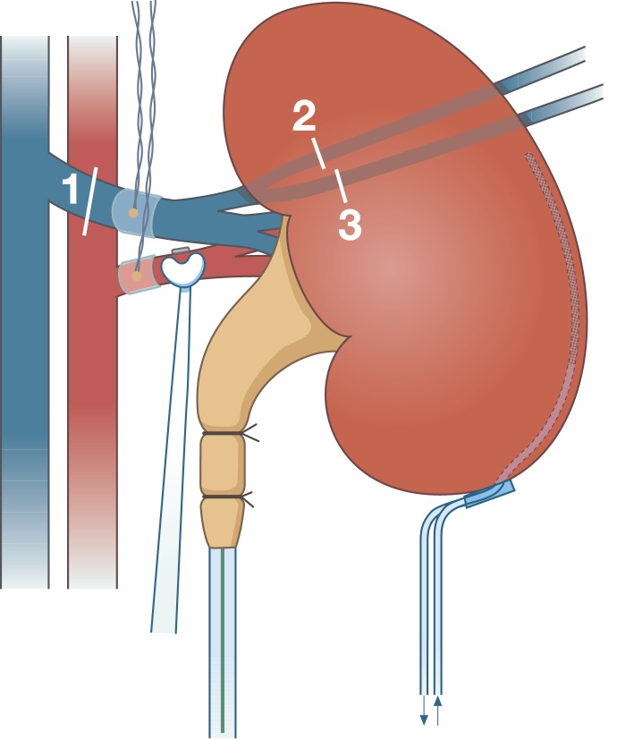
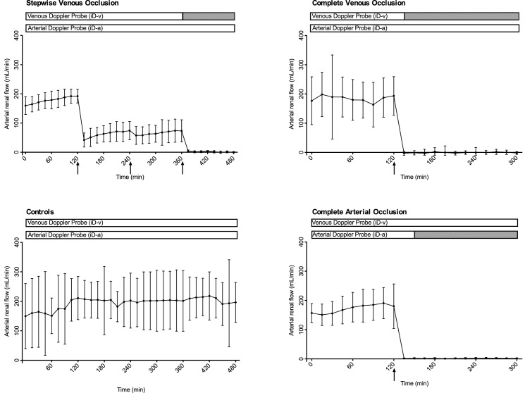
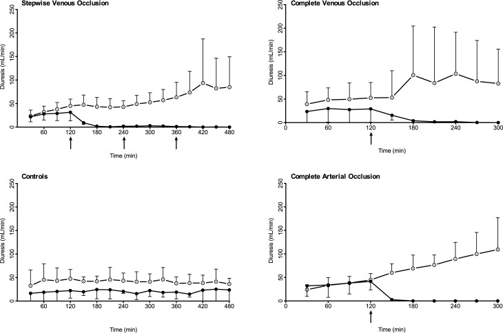
Methods: In 20 pigs, implantable Doppler probes were placed on the renal artery and vein and a microdialysis catheter was placed in the renal cortex. An arterial flowprobe served as gold standard. Following two-hour baseline measurements, the pigs were randomised to stepwise venous occlusion, complete venous occlusion, complete arterial occlusion or controls.
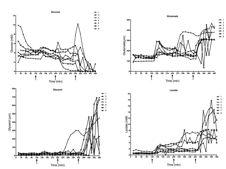
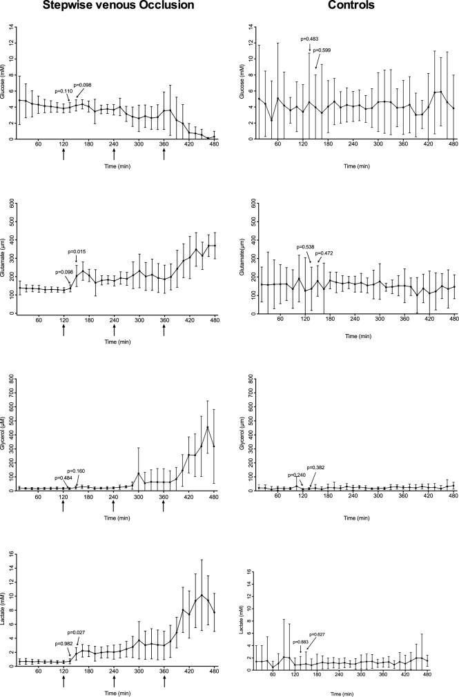
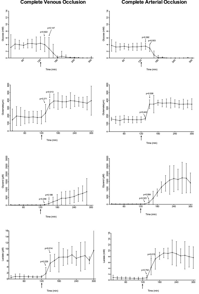
Results: All parameters were stable through baseline measurements. Glutamate and lactate measured by microdialysis increased significantly (p = 0.02 and p = 0.03 respectively) 30 minutes after a 2/3 (66%) reduction in renal blood flow. The implantable Doppler probe was not able to detect flow changes until there was total venous occlusion. Microdialysis detected changes in local metabolism after both arterial and venous occlusion; the implantable Doppler probe could only detect vascular occlusions on the vessel it was placed.
Conclusions: We developed a new model for stepwise renal venous blood flow occlusion. Furthermore, the first comparison of the implantable Doppler probe and microdialysis for detection of renal vascular occlusions was made. The implantable Doppler probe could only detect flow changes after a complete occlusion, whereas microdialysis detected changes earlier, and could detect both arterial and venous occlusion. Based on these results, the implantable Doppler probe for early detection of vascular occlusions cannot be recommended.
Conflict of interest statement
Figures
Similar articles
-
Detection of local metabolic changes after progressive and stepwise reduction of renal blood flow in pigs.Transplant Proc. 2009 Jan-Feb;41(1):44-8. doi: 10.1016/j.transproceed.2008.10.089. Transplant Proc. 2009. PMID: 19249471
-
Microdialysis for detection of renal ischemia after experimental renal transplantation.J Urol. 2009 Oct;182(4 Suppl):1854-9. doi: 10.1016/j.juro.2009.03.015. Epub 2009 Aug 19. J Urol. 2009. PMID: 19692033
-
Early detection of renal ischemia by in situ microdialysis: an experimental study.J Urol. 2008 Jan;179(1):371-5. doi: 10.1016/j.juro.2007.08.088. Epub 2007 Nov 19. J Urol. 2008. PMID: 18006006
-
Ultrasonography in kidney transplantation: values and new developments.Transplant Rev (Orlando). 2009 Oct;23(4):209-13. doi: 10.1016/j.trre.2009.06.003. Epub 2009 Aug 4. Transplant Rev (Orlando). 2009. PMID: 19654072 Review.
-
Vascular complications in the adult kidney transplant recipient.J Clin Ultrasound. 1992 Oct;20(8):517-27. doi: 10.1002/jcu.1870200805. J Clin Ultrasound. 1992. PMID: 1328321 Review.
Cited by
-
Involvement of the metabolic sensor GPR81 in cardiovascular control.JCI Insight. 2017 Oct 5;2(19):e92564. doi: 10.1172/jci.insight.92564. JCI Insight. 2017. PMID: 28978803 Free PMC article.
-
The functional activity of donor kidneys is negatively regulated by microribonucleic acid-451 in different perfusion methods to inhibit adenosine triphosphate metabolism and the proliferation of HK2 cells.Bioengineered. 2022 May;13(5):12706-12717. doi: 10.1080/21655979.2022.2068739. Bioengineered. 2022. PMID: 35603466 Free PMC article.
-
NIRS-based monitoring of kidney graft perfusion.PLoS One. 2020 Dec 2;15(12):e0243154. doi: 10.1371/journal.pone.0243154. eCollection 2020. PLoS One. 2020. PMID: 33264371 Free PMC article.
References
-
- Schmulder A, Gur E, Zaretski A. Eight-year experience of the Cook-Swartz Doppler in free-flap operations: Microsurgical and reexploration results with regard to a wide spectrum of surgeries. Microsurgery. 2011;31: 1–6. doi: 10.1002/micr.20816 - DOI - PubMed
-
- Sayegh MH, Carpenter CB. Transplantation 50 years later—progress, challenges, and promises. N Engl J Med. 2004;351: 2761–2766. doi: 10.1056/NEJMon043418 - DOI - PubMed
-
- Rozen W, Ang GG, McDonald A, Sivarajah G, Rahdon R, Acosta R, et al. Sutured Attachment of the Implantable Doppler Probe Cuff for Large or Complex Pedicles in Free Tissue Transfer. J reconstr Microsurg. 2010;27: 099–102. - PubMed
-
- Crane J, Hakim N. The use of an implantable Doppler flow probe in kidney transplantation: first report in the literature. Exp Clin Transplant. 2011;9: 118–120. - PubMed
-
- Ponticelli C, Moia M, Montagnino G. Renal allograft thrombosis. Nephrology Dialysis Transplantation. 2009;24: 1388–1393. - PubMed
MeSH terms
LinkOut - more resources
Full Text Sources
Other Literature Sources
Medical
