

Controlled delivery of tauroursodeoxycholic acid from biodegradable microspheres slows retinal degeneration and vision loss in P23H rats
- PMID: 28542454
- PMCID: PMC5444790
- DOI: 10.1371/journal.pone.0177998
Controlled delivery of tauroursodeoxycholic acid from biodegradable microspheres slows retinal degeneration and vision loss in P23H rats
Abstract
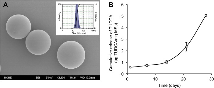
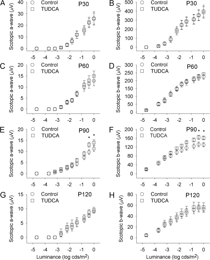
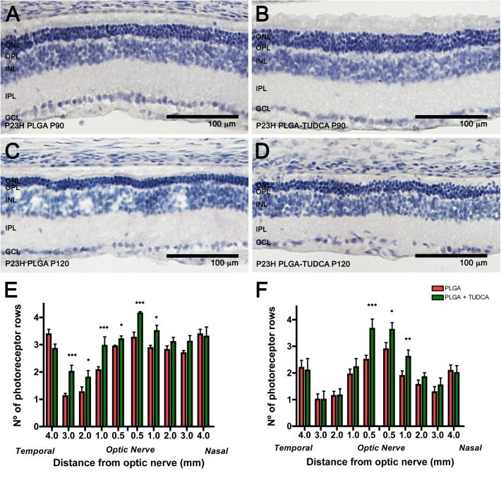
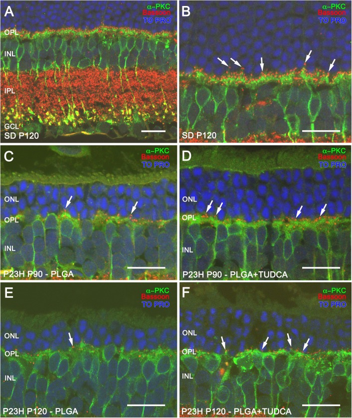
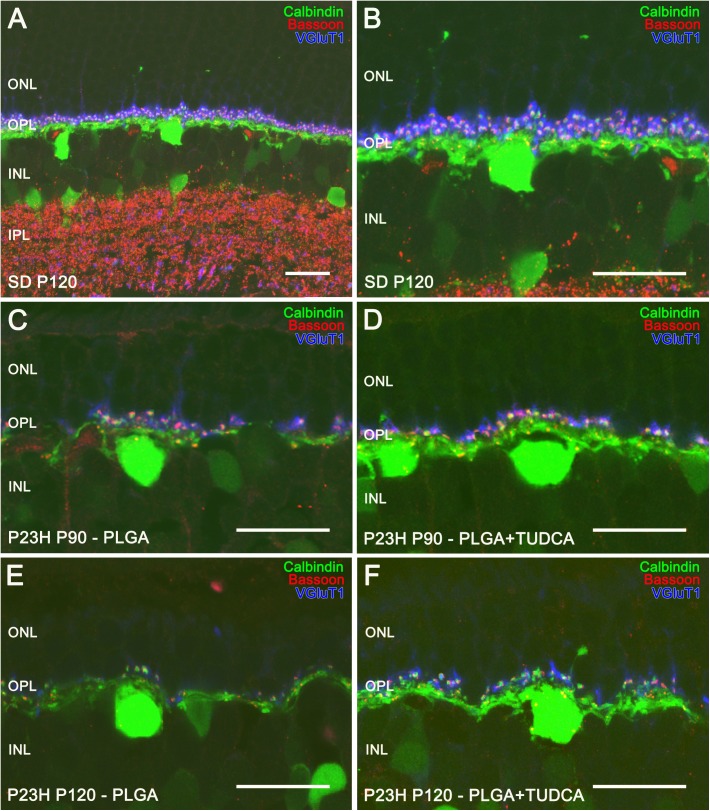
Successful drug therapies for treating ocular diseases require effective concentrations of neuroprotective compounds maintained over time at the site of action. The purpose of this work was to assess the efficacy of intravitreal controlled delivery of tauroursodeoxycholic acid (TUDCA) encapsulated in poly(D,L-lactic-co-glycolic acid) (PLGA) microspheres for the treatment of the retina in a rat model of retinitis pigmentosa. PLGA microspheres (MSs) containing TUDCA were produced by the O/W emulsion-solvent evaporation technique. Particle size and morphology were assessed by light scattering and scanning electronic microscopy, respectively. Homozygous P23H line 3 rats received a treatment of intravitreal injections of TUDCA-PLGA MSs. Retinal function was assessed by electroretinography at P30, P60, P90 and P120. The density, structure and synaptic contacts of retinal neurons were analyzed using immunofluorescence and confocal microscopy at P90 and P120. TUDCA-loaded PLGA MSs were spherical, with a smooth surface. The production yield was 78%, the MSs mean particle size was 23 μm and the drug loading resulted 12.5 ± 0.8 μg TUDCA/mg MSs. MSs were able to deliver the loaded active compound in a gradual and progressive manner over the 28-day in vitro release study. Scotopic electroretinografic responses showed increased ERG a- and b-wave amplitudes in TUDCA-PLGA-MSs-treated eyes as compared to those injected with unloaded PLGA particles. TUDCA-PLGA-MSs-treated eyes showed more photoreceptor rows than controls. The synaptic contacts of photoreceptors with bipolar and horizontal cells were also preserved in P23H rats treated with TUDCA-PLGA MSs. This work indicates that the slow and continuous delivery of TUDCA from PLGA-MSs has potential neuroprotective effects that could constitute a suitable therapy to prevent neurodegeneration and visual loss in retinitis pigmentosa.
Conflict of interest statement
Figures


Similar articles
-
Co-delivery of glial cell-derived neurotrophic factor (GDNF) and tauroursodeoxycholic acid (TUDCA) from PLGA microspheres: potential combination therapy for retinal diseases.Drug Deliv Transl Res. 2021 Apr;11(2):566-580. doi: 10.1007/s13346-021-00930-9. Epub 2021 Feb 27. Drug Deliv Transl Res. 2021. PMID: 33641047
-
Tauroursodeoxycholic acid prevents retinal degeneration in transgenic P23H rats.Invest Ophthalmol Vis Sci. 2011 Jul 1;52(8):4998-5008. doi: 10.1167/iovs.11-7496. Invest Ophthalmol Vis Sci. 2011. PMID: 21508111
-
Tolerance of high and low amounts of PLGA microspheres loaded with mineralocorticoid receptor antagonist in retinal target site.J Control Release. 2017 Nov 28;266:187-197. doi: 10.1016/j.jconrel.2017.09.029. Epub 2017 Sep 22. J Control Release. 2017. PMID: 28947395
-
Natural Compounds from Saffron and Bear Bile Prevent Vision Loss and Retinal Degeneration.Molecules. 2015 Jul 31;20(8):13875-93. doi: 10.3390/molecules200813875. Molecules. 2015. PMID: 26263962 Free PMC article. Review.
-
Neuroprotective Effect of Tauroursodeoxycholic Acid (TUDCA) on In Vitro and In Vivo Models of Retinal Disorders: A Systematic Review.Curr Neuropharmacol. 2024;22(8):1374-1390. doi: 10.2174/1570159X21666230907152207. Curr Neuropharmacol. 2024. PMID: 37691227 Free PMC article.
Cited by
-
Effects of Daily Melatonin Supplementation on Visual Loss, Circadian Rhythms, and Hepatic Oxidative Damage in a Rodent Model of Retinitis Pigmentosa.Antioxidants (Basel). 2021 Nov 22;10(11):1853. doi: 10.3390/antiox10111853. Antioxidants (Basel). 2021. PMID: 34829724 Free PMC article.
-
Short-term high-fat feeding exacerbates degeneration in retinitis pigmentosa by promoting retinal oxidative stress and inflammation.Proc Natl Acad Sci U S A. 2021 Oct 26;118(43):e2100566118. doi: 10.1073/pnas.2100566118. Proc Natl Acad Sci U S A. 2021. PMID: 34667124 Free PMC article.
-
Retinal Cell Protection in Ocular Excitotoxicity Diseases. Possible Alternatives Offered by Microparticulate Drug Delivery Systems and Future Prospects.Pharmaceutics. 2020 Jan 24;12(2):94. doi: 10.3390/pharmaceutics12020094. Pharmaceutics. 2020. PMID: 31991667 Free PMC article. Review.
-
Suppression of retinal degeneration by two novel ERAD ubiquitin E3 ligases SORDD1/2 in Drosophila.PLoS Genet. 2020 Nov 2;16(11):e1009172. doi: 10.1371/journal.pgen.1009172. eCollection 2020 Nov. PLoS Genet. 2020. PMID: 33137101 Free PMC article.
-
Co-delivery of glial cell-derived neurotrophic factor (GDNF) and tauroursodeoxycholic acid (TUDCA) from PLGA microspheres: potential combination therapy for retinal diseases.Drug Deliv Transl Res. 2021 Apr;11(2):566-580. doi: 10.1007/s13346-021-00930-9. Epub 2021 Feb 27. Drug Deliv Transl Res. 2021. PMID: 33641047
References
-
- Dryja TP, McGee TL, Reichel E, Hahn LB, Cowley GS, Yandell DW, et al. A point mutation of the rhodopsin gene in one form of retinitis pigmentosa. Nature. 1990;343(6256):364–6. Epub 1990/01/25. doi: 10.1038/343364a0 - DOI - PubMed
-
- Kaushal S, Khorana HG. Structure and function in rhodopsin. 7. Point mutations associated with autosomal dominant retinitis pigmentosa. Biochemistry. 1994;33(20):6121–8. Epub 1994/05/24. - PubMed
-
- Hartong DT, Berson EL, Dryja TP. Retinitis pigmentosa. Lancet. 2006;368(9549):1795–809. Epub 2006/11/23. doi: 10.1016/S0140-6736(06)69740-7 - DOI - PubMed
-
- Illing ME, Rajan RS, Bence NF, Kopito RR. A rhodopsin mutant linked to autosomal dominant retinitis pigmentosa is prone to aggregate and interacts with the ubiquitin proteasome system. J Biol Chem. 2002;277(37):34150–60. Epub 2002/07/02. M204955200 [pii]. doi: 10.1074/jbc.M204955200 - DOI - PubMed
MeSH terms
Substances
LinkOut - more resources
Full Text Sources
Other Literature Sources
