. 2017 May 24;12(5):e0178220.
doi: 10.1371/journal.pone.0178220. eCollection 2017.

A human monocytic NF-κB fluorescent reporter cell line for detection of microbial contaminants in biological samples

Affiliations

A human monocytic NF-κB fluorescent reporter cell line for detection of microbial contaminants in biological samples

Claire Battin et al. PLoS One. .

Abstract

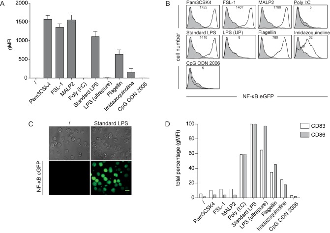
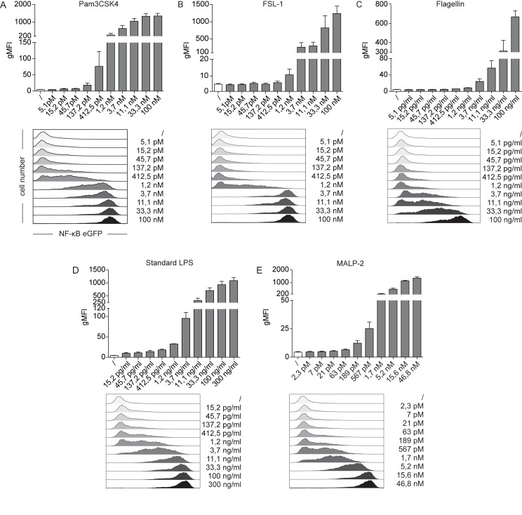
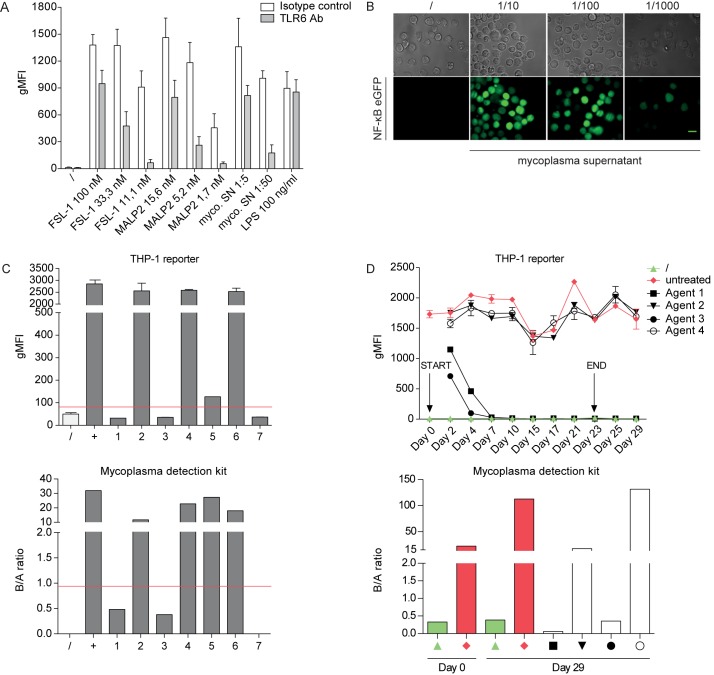
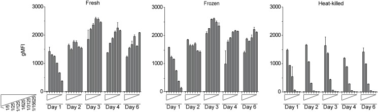
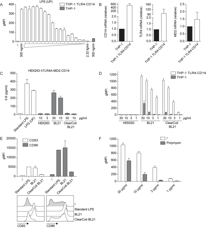
Sensing of pathogens by innate immune cells is essential for the initiation of appropriate immune responses. Toll-like receptors (TLRs), which are highly sensitive for various structurally and evolutionary conserved molecules derived from microbes have a prominent role in this process. TLR engagement results in the activation of the transcription factor NF-κB, which induces the expression of cytokines and other inflammatory mediators. The exquisite sensitivity of TLR signalling can be exploited for the detection of bacteria and microbial contaminants in tissue cultures and in protein preparations. Here we describe a cellular reporter system for the detection of TLR ligands in biological samples. The well-characterized human monocytic THP-1 cell line was chosen as host for an NF-ᴋB-inducible enhanced green fluorescent protein reporter gene. We studied the sensitivity of the resultant reporter cells for a variety of microbial components and observed a strong reactivity towards TLR1/2 and TLR2/6 ligands. Mycoplasma lipoproteins are potent TLR2/6 agonists and we demonstrate that our reporter cells can be used as reliable and robust detection system for mycoplasma contaminations in cell cultures. In addition, a TLR4-sensitive subline of our reporters was engineered, and probed with recombinant proteins expressed in different host systems. Bacterially expressed but not mammalian expressed proteins induced strong reporter activity. We also tested proteins expressed in an E. coli strain engineered to lack TLR4 agonists. Such preparations also induced reporter activation in THP-1 cells highlighting the importance of testing recombinant protein preparations for microbial contaminations beyond endotoxins. Our results demonstrate the usefulness of monocytic reporter cells for high-throughput screening for microbial contaminations in diverse biological samples, including tissue culture supernatants and recombinant protein preparations. Fluorescent reporter assays can be measured on standard flow cytometers and in contrast to established detection methods, like luciferase-based systems or Limulus Amebocyte Lysate tests, they do not require costly reagents.


Conflict of interest statement

Competing Interests: The authors have declared that no competing interests exist.

Figures

Fig 1
Fig 1. THP-1 NF-κB-eGFP reporter cells show a selective sensitivity towards TLR ligands.
(A) THP-1 NF-κB-eGFP cells were incubated with Pam3CSK4 (TLR1/2; 100 nM), FSL-1 (TLR2/6; 100 nM), MALP-2 (TLR2/6; 46,8 nM), Poly I:C (TLR3; 10 μg/ml), standard LPS (TLR2/4; 300 ng/ml), LPS ultrapure (UP) (TLR4; 300ng/ml), flagellin (TLR5; 100 ng/ml), imidazoquinoline (TLR7/8; 10 μg/ml) and CpG ODN 2006 (TLR9; 50 μg/ml). After 24 h, eGFP expression was assessed by flow cytometry. Bar graphs show geometric mean of fluorescence intensity (gMFI). Mean and SE were calculated from triplicates of five independently performed experiments (n = 5). (B) Representative flow cytometry histograms of reporter gene expression in THP-1 NF-κB-eGFP cells as described in A. Open histograms: TLR-activated reporter cells; filled histograms: unstimulated reporter cells. Numbers show gMFI. (C) Fluorescent microscopy images of reporter cells activated with standard LPS (3 μg/ml) for 24 h (right panel). Unstimulated cells served as negative control (left panel). Bright field images are shown for comparison (top row). Scale bar: 10 μm. (D) Immature human monocyte-derived DCs were incubated with various TLR ligands (used at the same concentrations as in A) for 24 h or were left untreated. Expression of maturation markers CD83 and CD86 was assessed by flow cytometry. Bar graphs show total percentage of gMFI normalized to standard LPS.
Fig 2
Fig 2. Dose-dependent response of THP-1 NF-κB-eGFP reporter cells towards specific TLR ligands.
(A-E) THP-1 NF-κB-eGFP cells were incubated with increasing concentrations of Pam3CSK4, FSL-1, Flagellin, standard LPS and MALP-2 as indicated. Untreated cells served as control. After 24 h, induction of NF-κB-driven eGFP was measured by flow cytometry. Bar graphs show geometric mean of fluorescence intensity (gMFI, top panels). Mean and SE were calculated from triplicates of three independently performed experiments (n = 3). Flow cytometry histograms of a representative experiment are shown for comparison (bottom panels). Open histograms: control cells; filled histograms: TLR-activated reporter cells.
Fig 3
Fig 3. THP-1 NF-κB-eGFP reporter cells are highly sensitive towards mycoplasma lipoproteins through engagement of TLR2/6.
(A) THP-1 reporter cells pre-treated with a blocking TLR6 antibody or isotype control (30 min; both used at 5 μg/mL) were incubated with the indicated concentrations of the TLR2/6 ligands FSL-1 and MALP-2 or supernatants derived from mycoplasma infected cell cultures for 24 h. Standard LPS served as a negative control. NF-κB-driven eGFP expression was assessed by flow cytometry. Bar graphs show geometric mean of fluorescence intensity (gMFI). Mean and SE were calculated from triplicates of three independently performed experiments (n = 3). (B) Fluorescent microscopy images of THP-1 reporter cells stimulated for 24 h with different dilutions of supernatants derived from mycoplasma infected cell cultures. Unstimulated cells served as negative control (left panel). Bright field images are shown for comparison (top row). Scale bar: 10 μm. (C) THP-1 NF-κB-eGFP reporter cells and a commercially available mycoplasma detection kit (MycoAlert) were probed with tissue culture supernatants from different cell sources and species: (1) mouse tail cells, (2) human mesotheliom, (3) human melanoma brain metastasis-derived cell line YDFR, (4) human LN229 glioblastoma, (5) human ovarian cancer cells, (6) COS-7 cell line and (7) human skin fibroblasts. Samples above red line are scored as positive. For the Mycoalert detection system the B/A ratio represents the ratio of the luminescence signals measured at two different time points (reading A and B). For THP-1 reporter assay mean and SE were calculated from duplicates. (D) K562 cells were infected with mycoplasma and then subjected to treatment regimens using commercially available mycoplasma removal agents (see Material and Methods). Supernatants were collected at various time points throughout the treatment course (day 2, 4, 7, 10, 15, 17, 21, 23, 25 and 29) and tested with the THP-1 NF-κB-eGFP reporter cells (upper panel). Indicated samples were also tested using the MycoAlert kit (lower panel).
Fig 4
Fig 4. THP-1 NF-κB-eGFP reporters can detect the presence of mycoplasma in heat-denatured and cryo-preserved samples.
THP-1 reporter cells were incubated with fresh (left panel), frozen (middle panel) or 95°C heat-inactivated (right panel) mycoplasma-containing tissue culture supernatants used at the indicated dilutions. NF-κB-driven eGFP expression was measured at day 1, 2, 3, 4 and 6 after the onset of the assay by flow cytometry. Bar graphs show geometric mean of fluorescence intensity (gMFI). Mean and SE were calculated from duplicates.
Fig 5
Fig 5. Use of THP-1 TLR4-CD14 NF-κB-eGFP reporter cells for the detection of microbial contaminations in recombinant protein preparations.
(A) THP-1 TLR4-CD14 NF-κB-eGFP reporter cells were incubated with the indicated serial dilutions of ultrapure LPS for 24 h. NF-κB-driven eGFP expression was assessed by flow cytometry. Bar graphs show geometric mean of fluorescence intensity (gMFI). eGFP expression of the parental THP-1 reporter cells treated with 300 ng/ml ultrapure LPS is shown for comparison (grey bar). (B) mRNA expression of TLR4, CD14 and MD2 was measured in parental and TLR4-CD14 THP-1 reporter cells by real-time qPCR. Data are presented as mean ± SEM of expression values normalized against the housekeeping gene GAPDH (n = 4). (C) Recombinant human split product C4dg produced in three different expression systems (HEK293-6E, standard E. coli BL21 and E. coli ClearColi BL21) was tested for TLR4-agonist contaminations using HEK293 hTLR4A-MD2-CD14 cells. Following 24 h of incubation the IL-8 content in the culture supernatants was measured by ELISA. (D) Parental and TLR4-CD14 THP-1 reporters were incubated with the protein preparations described in (C) and NF-κB-driven eGFP expression was assessed by flow cytometry 24 h later. Bar graphs show geometric mean of fluorescence intensity (gMFI). Mean and SE were calculated from duplicates of four independently performed experiments (n = 4) (E) Immature human moDCs were incubated with standard LPS, E. coli BL21 or E. coli ClearColi BL21 expressed C4dg protein at the indicated concentrations for 24 h or were left untreated. Expression of maturation markers CD83 and CD86 was assessed by flow cytometry. (F) E. coli ClearColi BL21-expressed C4dg protein was subjected to a single round of bulk chromatography using a polymyxin resin (see Material and Methods). Samples before and after chromatography were tested using the THP-1 TLR4-CD14 reporters. Mean and SE were calculated from duplicates of four independently performed experiments (n = 4).

References

    1. Cooper JF, Levin J, Wagner HN Jr. Quantitative comparison of in vitro and in vivo methods for the detection of endotoxin. The Journal of laboratory and clinical medicine. 1971;78(1):138–48. - PubMed
    1. Drexler HG, Uphoff CC. Mycoplasma contamination of cell cultures: Incidence, sources, effects, detection, elimination, prevention. Cytotechnology. 2002;39(2):75–90. doi: 10.1023/A:1022913015916 - DOI - PMC - PubMed
    1. Lemaitre B, Nicolas E, Michaut L, Reichhart JM, Hoffmann JA. The dorsoventral regulatory gene cassette spatzle/Toll/cactus controls the potent antifungal response in Drosophila adults. Cell. 1996;86(6):973–83. - PubMed
    1. Gay NJ, Keith FJ. Drosophila Toll and IL-1 receptor. Nature. 1991;351(6325):355–6. doi: 10.1038/351355b0 - DOI - PubMed
    1. Medzhitov R, Preston-Hurlburt P, Janeway CA Jr. A human homologue of the Drosophila Toll protein signals activation of adaptive immunity. Nature. 1997;388(6640):394–7. doi: 10.1038/41131 - DOI - PubMed

MeSH terms