. 2017 May 24;12(5):e0178370.
doi: 10.1371/journal.pone.0178370. eCollection 2017.

Functional interaction between co-expressed MAGE-A proteins

Affiliations

Functional interaction between co-expressed MAGE-A proteins

Julieta E Laiseca et al. PLoS One. .

Abstract

MAGE-A (Melanoma Antigen Genes-A) are tumor-associated proteins with expression in a broad spectrum of human tumors and normal germ cells. MAGE-A gene expression and function are being increasingly investigated to better understand the mechanisms by which MAGE proteins collaborate in tumorigenesis and whether their detection could be useful for disease prognosis purposes. Alterations in epigenetic mechanisms involved in MAGE gene silencing cause their frequent co-expression in tumor cells. Here, we have analyzed the effect of MAGE-A gene co-expression and our results suggest that MageA6 can potentiate the androgen receptor (AR) co-activation function of MageA11. Database search confirmed that MageA11 and MageA6 are co-expressed in human prostate cancer samples. We demonstrate that MageA6 and MageA11 form a protein complex resulting in the stabilization of MageA11 and consequently the enhancement of AR activity. The mechanism involves association of the Mage A6-MHD domain to MageA11, prevention of MageA11 ubiquitinylation on lysines 240 and 245 and decreased proteasome-dependent degradation. We experimentally demonstrate here for the first time that two MAGE-A proteins can act together in a non-redundant way to potentiate a specific oncogenic function. Overall, our results highlight the complexity of the MAGE gene networking in regulating cancer cell behavior.


Conflict of interest statement

Competing Interests: The authors have declared that no competing interests exist.

Figures

Fig 1
Fig 1. MageA6 enhances MageA11-dependent AR transcriptional activity.
(A) Reporter gene assay for GR, MR and AR activity using specific gene-reporter in the presence or absence of MageA6 or MageA11 expression. Cells were treated with dexamethasone (Dx), Aldosterone (Aldo) or dihidrotestosterone (DHT) for 24 h prior to harvesting. The assay was performed in HEK293T cells. ev, empty vector. (B) Similar to A but combining MageA11 and MageA6 expression as indicated. (C) Determination of PSA mRNA levels through RT-qPCR. LNCaP cells were transfected with a siRNA control (siC) or a siRNA to silence MageA6 expression (siA6/2). DHT was added to cells as indicated. PSA mRNA levels were normalized to GAPDH mRNA levels. Error bars indicate mean S.D. Student’s t test was used for statistical analysis. * p < 0.05. ** p < 0.001.
Fig 2
Fig 2. MageA6 and MageA11 co-expression in prostate cancer.
Analysis of cBioPortal Cancer Genomics data sets form Prostate Adenocarcinoma and Testicular Germ Cell Cancer (see Material and Methods) as indicated in the two main columns. Gene overexpression was calculated by Z-score, defined as the relative expression of an individual gene to the gene’s expression distribution in a reference population. The indicated percent of over-expression refers to the number of samples over-expressing a given gene over the total of samples. Dot-plot graphics shows the correlation between MageA6 and MageA11 gene expression. Insets indicate Pearson and Spearman correlation scores.
Fig 3
Fig 3. MageA6 associates with MageA11 in cells.
(A) Co-immunoprecipitation of MageA11 and Mage-A proteins from LNCaP cells. Specific anti-MageA11 antibody was used to immunoprecipitate MageA11. Western blot was performed using a pan-MAGE-A antibody. Anti-GFP was used as IP control antibody. (B) Co-Immunoprecipitation of Flag-MageA11 and HA-MageA6 form HEK293T cells. Immunoprecipitated protein complexes obtained with the anti-Flag antibody (IP anti-Flag) were assessed by Western blot and probed with the indicated antibodies. TL, total lysates. (C) Similar to A but using Flag-MageA11 and GFP-MHD-MageA6. (D) Upper panel: Reporter gene assay for AR activity in HEK293T cells using the PSA-Luc reporter construct and the plasmid encoding AR. MageA11, MageA6, MageA2 and MHD-MageA6 or MHD-MageA2 were co-transfected as indicated in the figure. Cells were treated with DHT 24 h prior to harvest. ev, empty vector. Bottom panel: A sketch of Mage-A protein domains and their expected MW. Error bars indicate mean S.D. Student’s t test was used for statistical analysis. * p < 0.001; ** Immunoglobulin heavy chain. *** Immunoglobulin light chain. Triangles show the corresponding protein band and dashes mark the MW.
Fig 4
Fig 4. MageA6 expression increases MageA11 protein levels.
(A)Western blot showing Flag-MageA11 when co-expressed with increasing quantities of HA-MageA6 (0, 250, 500, 1000 and 1500 ng). GFP expression is the internal control. Membrane was probed with the indicated antibodies. (B) Western blot showing Flag-MageA11 when co-expressed with increasing quantities of HA-MHD-MageA6 (0, 250, 500, 1000 and 1500 ng). GFP expression is the internal control. Membrane was probed with the indicated antibodies. (C) Western blot showing Flag-MageA11 when co-expressed with increasing quantities of HA-MageA2 (0, 250, 500, 1000 and 1500 ng). GFP expression is the internal control. Membrane was probed with the indicated antibodies. (D) Western blot of LNCaP cells silenced (siA6/2) or not (siC) for MageA6 expression. Anti-pan MAGE-A antibody (6C1, Santa Cruz) was used to detect Mage-A proteins. 65KDa band corresponds to MageA11 while 45KDa band could correspond to different Mage-A proteins. GAPDH was used as loading control. (E)Left panel: Western Blot showing the endogenous levels of MageA11 in LNCaP stably expressing MageA6 (A6) or empty vector (EV). Extracts of HEK293T cells transfected with Flag-MageA11 (F-A11), HA-MageA6 (HA-A6) or empty vector (EV) were used as controls. MAGE-A detection was performed with anti-pan MAGE antibody (6C1, Santa Cruz). 65KDa band corresponds to MageA11 while 45KDa band could correspond to different Mage-A proteins. The observed increment in 45KDa band in LNCaP-A6 is caused by MageA6 stable expression. Right panel: quantification of MAGE-A11 vs β-tubulin band intensity corresponding to Fig 4E, lanes 4 and 5. (F) RT-qPCR for the determination of MageA11 mRNA levels in LNCaP-A6 (A6) and LNCaP-EV (EV). MageA11 mRNA was normalized to GAPDH mRNA levels. (G) RT-qPCR for the determination of PSA mRNA levels in LNCaP-A6 (A6) and LNCaP-EV (EV). PSA mRNA was normalized to GAPDH mRNA levels. Error bars indicate mean S.D. Student’s t test was used for statistical analysis. ** p < 0.001. * unspecific band. Triangles show the corresponding protein band and dashes mark the MW.
Fig 5
Fig 5. Proteasome-dependent increase of MageA11 by MageA6 expression.
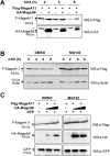
(A)Western blot showing Flag-MageA11 alone or when co-expressed with HA-MageA6 after 0, 3 and 6 hours of cycloheximide (CHX) treatment. The membrane was probed with the indicated antibodies. (B) Western blot showing Flag-MageA11 expression in the presence of MG132 (1,5uM for 20h) or absence of MG132 (DMSO) after 0, 2, 4 and 6 hours of cycloheximide (CHX) treatment. The membrane was probed with the indicated antibodies. (C) Western blot showing Flag-MageA11 levels alone or when co-expressed with HA-MageA6 in the presence of MG132 or absence of MG132 (DMSO). GFP expression is the internal control. Membrane was probed with the indicated antibodies. *Unspecific band. Triangles show the corresponding protein band and dashes mark the MW.
Fig 6
Fig 6. MageA6 interaction with MageA11 prevents MageA11 ubiquitinylation.
(A) In vivo ubiquitinylation assay in HEK293T cells transfected with Flag-MageA11 and HA-ubiquitin with or without HA-MageA6. Cells were treated with MG132 (10μM) to favor accumulation of ubiquitinylated proteins. Flag-MageA11was immunoprecipitated with anti-Flag antibody (IP anti-Flag). Immunoprecipitated material was assessed by Western blot and probed with the anti-HA antibody. (B) In vivo ubiquitinylation assay in HEK293T cells transfected with Flag-MageA11 or Flag-MageA11-2KA (Flag-MageA11K240A;K245A) and HA-ubiquitin. Cells were treated with MG132 (10μM) to favor accumulation of ubiquitinylated proteins. Flag-MageA11 was immunoprecipitated with anti-Flag antibody (IP anti-Flag). Immunoprecipitated material was assessed by Western blot and probed with the anti-HA antibody. (C) Western blot showing Flag-MageA11-wt or Flag-MageA11-2KA protein levels of in HEK293T cells transfected with equal amounts of vector DNA as indicated. The day after transfection, cells were incubated with cycloheximide (CHX) for 0, 2, 4, 6 and 8 hours. GFP is an internal control. Membrane was probed with the indicated antibodies. (D) Western blot showing Flag-MageA11-wt or Flag-MageA11-2KA protein levels when co-expressed with increased amounts (0 ug, 1 ug and 2 ug) of HA-MageA6 expressing vector. GFP is an internal control. Membrane was probed with the indicated antibodies. (E) Quantification of Flag-MAGE-A11 or Flag-MageA11-2KA band intensity corresponding to Fig 6D and normalized to GFP. * Unspecific band. ** Immunoglobulin heavy chain. Triangles show the corresponding protein band and dashes mark the MW.

References

    1. Simpson AJG, Caballero OL, Jungbluth A, Chen Y-T, Old LJ. Cancer/testis antigens, gametogenesis and cancer. Nat Rev Cancer. 2005;5: 615–625. doi: 10.1038/nrc1669 - DOI - PubMed
    1. van der Bruggen P, Traversari C, Chomez P, Lurquin C, De Plaen E, Van den Eynde B, et al. A gene encoding an antigen recognized by cytolytic T lymphocytes on a human melanoma. Science. 1991;254: 1643–7. - PubMed
    1. Newman JA, Cooper CDO, Roos AK, Aitkenhead H, Oppermann UCT, Cho HJ, et al. Structures of Two Melanoma-Associated Antigens Suggest Allosteric Regulation of Effector Binding. PLoS One. 2016;11: e0148762 doi: 10.1371/journal.pone.0148762 - DOI - PMC - PubMed
    1. Meek DW, Marcar L. MAGE-A antigens as targets in tumour therapy. Cancer Lett. Elsevier Ireland Ltd; 2012;324: 126–32. doi: 10.1016/j.canlet.2012.05.011 - DOI - PubMed
    1. Ladelfa MF, Peche LY, Toledo MF, Laiseca JE, Schneider C, Monte M. Tumor-specific MAGE proteins as regulators of p53 function. Cancer Lett. 2012;325: 11–17. doi: 10.1016/j.canlet.2012.05.031 - DOI - PubMed

MeSH terms

Supplementary concepts