

The structural basis of a high affinity ATP binding ε subunit from a bacterial ATP synthase
- PMID: 28542497
- PMCID: PMC5436830
- DOI: 10.1371/journal.pone.0177907
The structural basis of a high affinity ATP binding ε subunit from a bacterial ATP synthase
Abstract
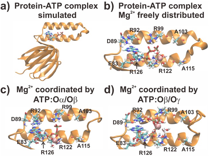
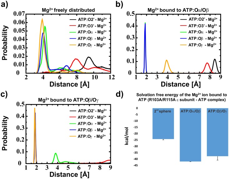
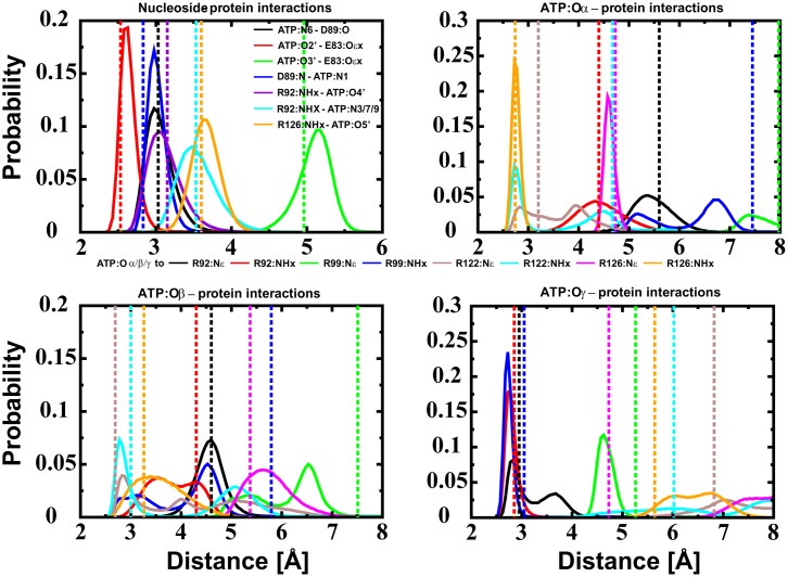
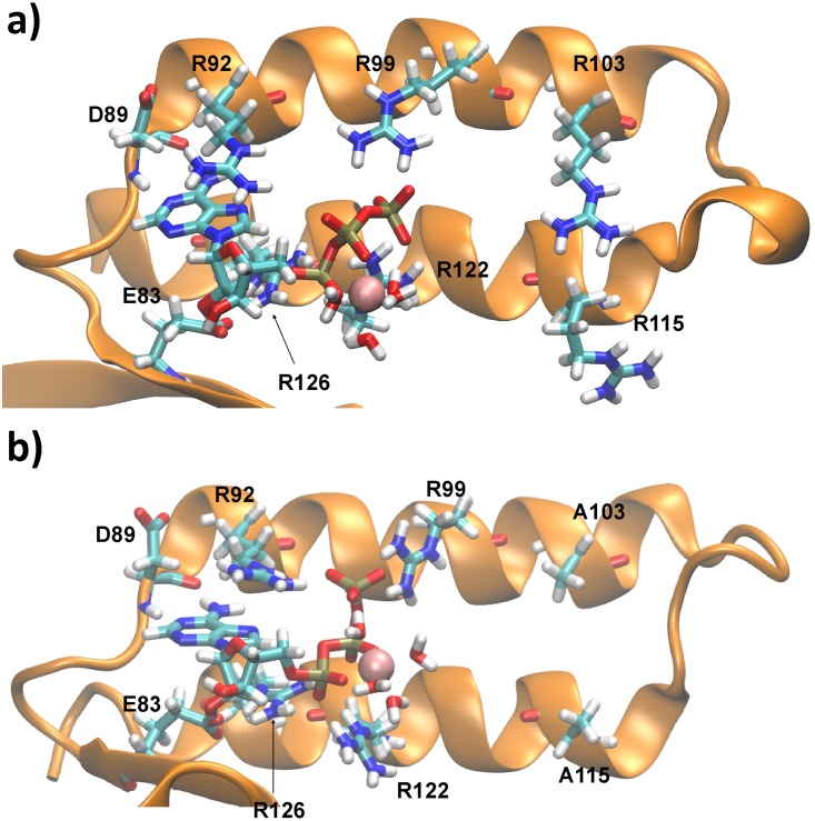
The ε subunit from bacterial ATP synthases functions as an ATP sensor, preventing ATPase activity when the ATP concentration in bacterial cells crosses a certain threshold. The R103A/R115A double mutant of the ε subunit from thermophilic Bacillus PS3 has been shown to bind ATP two orders of magnitude stronger than the wild type protein. We use molecular dynamics simulations and free energy calculations to derive the structural basis of the high affinity ATP binding to the R103A/R115A double mutant. Our results suggest that the double mutant is stabilized by an enhanced hydrogen-bond network and fewer repulsive contacts in the ligand binding site. The inferred structural basis of the high affinity mutant may help to design novel nucleotide sensors based on the ε subunit from bacterial ATP synthases.
Conflict of interest statement
Figures
Similar articles
-
Single mutations in the ε subunit from thermophilic Bacillus PS3 generate a high binding affinity site for ATP.PeerJ. 2018 Sep 5;6:e5505. doi: 10.7717/peerj.5505. eCollection 2018. PeerJ. 2018. PMID: 30202650 Free PMC article.
-
The Molecular Basis for Purine Binding Selectivity in the Bacterial ATP Synthase ϵ Subunit.Chembiochem. 2020 Nov 16;21(22):3249-3254. doi: 10.1002/cbic.202000291. Epub 2020 Aug 4. Chembiochem. 2020. PMID: 32608105
-
High affinity nucleotide-binding mutant of the ε subunit of thermophilic F1-ATPase.Biochem Biophys Res Commun. 2016 Jan 22;469(4):1129-32. doi: 10.1016/j.bbrc.2015.12.121. Epub 2015 Dec 30. Biochem Biophys Res Commun. 2016. PMID: 26746006
-
Rotating proton pumping ATPases: subunit/subunit interactions and thermodynamics.IUBMB Life. 2013 Mar;65(3):247-54. doi: 10.1002/iub.1134. IUBMB Life. 2013. PMID: 23441040 Review.
-
Structure and mechanism of Escherichia coli RecA ATPase.Mol Microbiol. 2005 Oct;58(2):358-66. doi: 10.1111/j.1365-2958.2005.04876.x. Mol Microbiol. 2005. PMID: 16194225 Review.
Cited by
-
The advancement of biosensor design and construction utilizing biomolecular motors.Synth Syst Biotechnol. 2025 Feb 18;10(2):543-554. doi: 10.1016/j.synbio.2025.02.007. eCollection 2025 Jun. Synth Syst Biotechnol. 2025. PMID: 40092161 Free PMC article. Review.
-
ATP binding by an F1Fo ATP synthase ε subunit is pH dependent, suggesting a diversity of ε subunit functional regulation in bacteria.Front Mol Biosci. 2023 Feb 27;10:1059673. doi: 10.3389/fmolb.2023.1059673. eCollection 2023. Front Mol Biosci. 2023. PMID: 36923639 Free PMC article.
-
Control of rotation of the F1FO-ATP synthase nanomotor by an inhibitory α-helix from unfolded ε or intrinsically disordered ζ and IF1 proteins.J Bioenerg Biomembr. 2018 Oct;50(5):403-424. doi: 10.1007/s10863-018-9773-9. Epub 2018 Sep 28. J Bioenerg Biomembr. 2018. PMID: 30267331 Review.
-
Changes within the central stalk of E. coli F1Fo ATP synthase observed after addition of ATP.Commun Biol. 2023 Jan 11;6(1):26. doi: 10.1038/s42003-023-04414-z. Commun Biol. 2023. PMID: 36631659 Free PMC article.
-
Single mutations in the ε subunit from thermophilic Bacillus PS3 generate a high binding affinity site for ATP.PeerJ. 2018 Sep 5;6:e5505. doi: 10.7717/peerj.5505. eCollection 2018. PeerJ. 2018. PMID: 30202650 Free PMC article.
References
-
- Mitchell P. Coupling of phosphorylation to electron and hydrogen transfer by a chemi-osmotic type of mechanism. Nature. 1961;191: 144–8. Available: http://www.ncbi.nlm.nih.gov/pubmed/13771349 - PubMed
MeSH terms
Substances
LinkOut - more resources
Full Text Sources
Other Literature Sources
