

Ectopically expressed Slc34a2a sense-antisense transcripts cause a cerebellar phenotype in zebrafish embryos depending on RNA complementarity and Dicer
- PMID: 28542524
- PMCID: PMC5436864
- DOI: 10.1371/journal.pone.0178219
Ectopically expressed Slc34a2a sense-antisense transcripts cause a cerebellar phenotype in zebrafish embryos depending on RNA complementarity and Dicer
Abstract
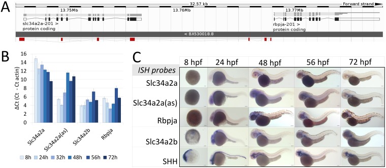
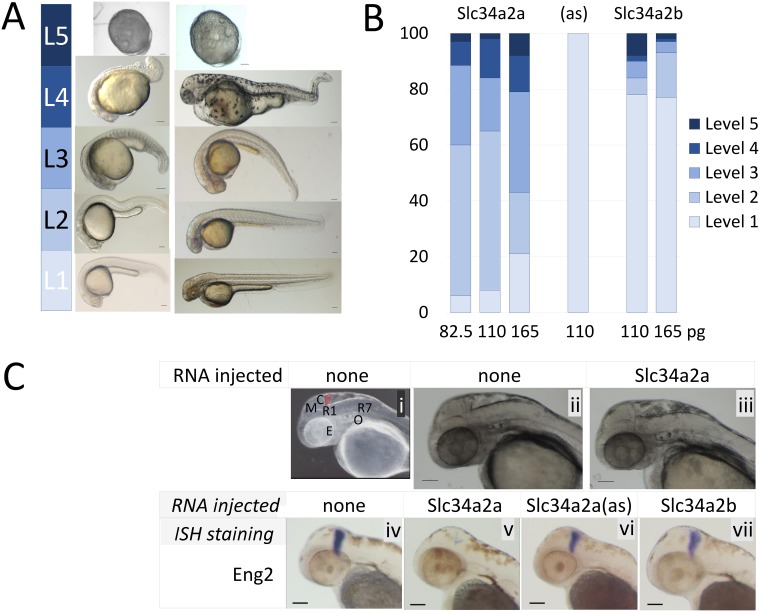
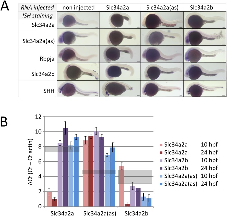
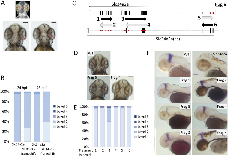
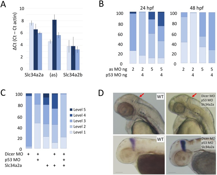
Natural antisense transcripts (NATs) are complementary to protein coding genes and potentially regulate their expression. Despite widespread occurrence of NATs in the genomes of higher eukaryotes, their biological role and mechanism of action is poorly understood. Zebrafish embryos offer a unique model system to study sense-antisense transcript interplay at whole organism level. Here, we investigate putative antisense transcript-mediated mechanisms by ectopically co-expressing the complementary transcripts during early zebrafish development. In zebrafish the gene Slc34a2a (Na-phosphate transporter) is bi-directionally transcribed, the NAT predominantly during early development up to 48 hours after fertilization. Declining levels of the NAT, Slc34a2a(as), coincide with an increase of the sense transcript. At that time, sense and antisense transcripts co-localize in the endoderm at near equal amounts. Ectopic expression of the sense transcript during embryogenesis leads to specific failure to develop a cerebellum. The defect is RNA-mediated and dependent on sense-antisense complementarity. Overexpression of a Slc34a2a paralogue (Slc34a2b) or the NAT itself had no phenotypic consequences. Knockdown of Dicer rescued the brain defect suggesting that RNA interference is required to mediate the phenotype. Our results corroborate previous reports of Slc34a2a-related endo-siRNAs in two days old zebrafish embryos and emphasize the importance of coordinated expression of sense-antisense transcripts. Our findings suggest that RNAi is involved in gene regulation by certain natural antisense RNAs.
Conflict of interest statement
Figures
Similar articles
-
Processing of naturally occurring sense/antisense transcripts of the vertebrate Slc34a gene into short RNAs.Physiol Genomics. 2008 Jun 12;34(1):95-100. doi: 10.1152/physiolgenomics.00004.2008. Epub 2008 Apr 15. Physiol Genomics. 2008. PMID: 18413783
-
Small RNAs and the regulation of cis-natural antisense transcripts in Arabidopsis.BMC Mol Biol. 2008 Jan 14;9:6. doi: 10.1186/1471-2199-9-6. BMC Mol Biol. 2008. PMID: 18194570 Free PMC article.
-
Strand selective generation of endo-siRNAs from the Na/phosphate transporter gene Slc34a1 in murine tissues.Nucleic Acids Res. 2009 Apr;37(7):2274-82. doi: 10.1093/nar/gkp088. Epub 2009 Feb 23. Nucleic Acids Res. 2009. PMID: 19237395 Free PMC article.
-
Regulation of the NPT gene by a naturally occurring antisense transcript.Cell Biochem Biophys. 2002;36(2-3):241-52. doi: 10.1385/CBB:36:2-3:241. Cell Biochem Biophys. 2002. PMID: 12139410 Review.
-
What do natural antisense transcripts regulate?RNA Biol. 2009 Jan-Mar;6(1):43-8. doi: 10.4161/rna.6.1.7568. Epub 2009 Jan 2. RNA Biol. 2009. PMID: 19098462 Review.
Cited by
-
Type II Na+-phosphate Cotransporters and Phosphate Balance in Teleost Fish.Pflugers Arch. 2019 Jan;471(1):193-212. doi: 10.1007/s00424-018-2239-4. Epub 2018 Dec 12. Pflugers Arch. 2019. PMID: 30542786 Review.
-
Mechanisms of Antisense Transcription Initiation with Implications in Gene Expression, Genomic Integrity and Disease Pathogenesis.Noncoding RNA. 2019 Jan 21;5(1):11. doi: 10.3390/ncrna5010011. Noncoding RNA. 2019. PMID: 30669611 Free PMC article. Review.
-
Endogenous Double-Stranded RNA.Noncoding RNA. 2021 Feb 19;7(1):15. doi: 10.3390/ncrna7010015. Noncoding RNA. 2021. PMID: 33669629 Free PMC article. Review.
-
A cautionary tale of sense-antisense gene pairs: independent regulation despite inverse correlation of expression.Nucleic Acids Res. 2017 Dec 1;45(21):12496-12508. doi: 10.1093/nar/gkx952. Nucleic Acids Res. 2017. PMID: 29059299 Free PMC article.
-
Genome-wide identification of antisense lncRNAs and their association with susceptibility to Flavobacterium psychrophilum in rainbow trout.Front Immunol. 2022 Dec 6;13:1050722. doi: 10.3389/fimmu.2022.1050722. eCollection 2022. Front Immunol. 2022. PMID: 36561762 Free PMC article.
References
-
- Werner A, Carlile M, Swan D. What do natural antisense transcripts regulate? RNA Biol. 2009;6(1):43–8. Epub 2008/12/23. . - PubMed
MeSH terms
Substances
Grants and funding
LinkOut - more resources
Full Text Sources
Other Literature Sources
Molecular Biology Databases
