. 2017 May 25;13(5):e1006346.
doi: 10.1371/journal.ppat.1006346. eCollection 2017 May.

Varicella zoster virus glycoprotein C increases chemokine-mediated leukocyte migration

Affiliations

Varicella zoster virus glycoprotein C increases chemokine-mediated leukocyte migration

Víctor González-Motos et al. PLoS Pathog. .

Abstract

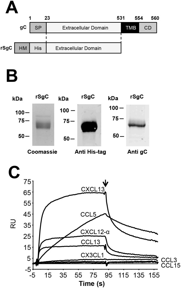
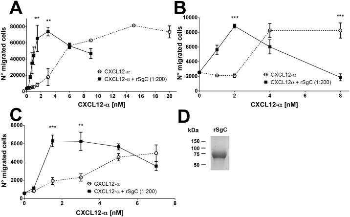
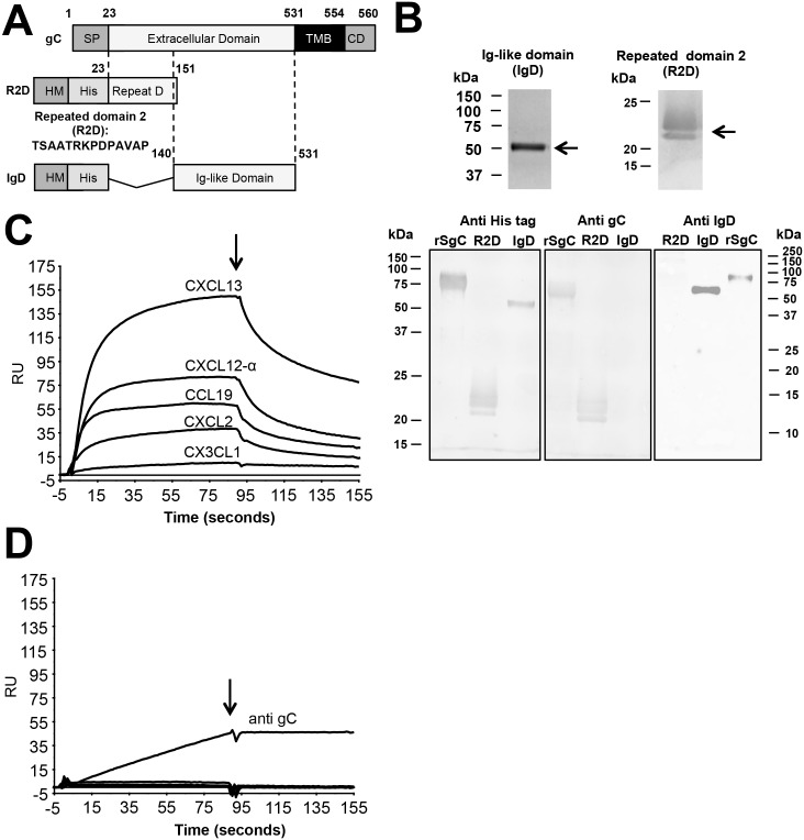
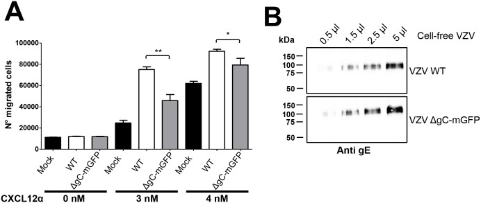
Varicella zoster virus (VZV) is a highly prevalent human pathogen that establishes latency in neurons of the peripheral nervous system. Primary infection causes varicella whereas reactivation results in zoster, which is often followed by chronic pain in adults. Following infection of epithelial cells in the respiratory tract, VZV spreads within the host by hijacking leukocytes, including T cells, in the tonsils and other regional lymph nodes, and modifying their activity. In spite of its importance in pathogenesis, the mechanism of dissemination remains poorly understood. Here we addressed the influence of VZV on leukocyte migration and found that the purified recombinant soluble ectodomain of VZV glycoprotein C (rSgC) binds chemokines with high affinity. Functional experiments show that VZV rSgC potentiates chemokine activity, enhancing the migration of monocyte and T cell lines and, most importantly, human tonsillar leukocytes at low chemokine concentrations. Binding and potentiation of chemokine activity occurs through the C-terminal part of gC ectodomain, containing predicted immunoglobulin-like domains. The mechanism of action of VZV rSgC requires interaction with the chemokine and signalling through the chemokine receptor. Finally, we show that VZV viral particles enhance chemokine-dependent T cell migration and that gC is partially required for this activity. We propose that VZV gC activity facilitates the recruitment and subsequent infection of leukocytes and thereby enhances VZV systemic dissemination in humans.


Conflict of interest statement

AEIP's affiliation is NovImmune, Geneva, Switzerland. The authors have declared that no competing interests exist.

Figures

Fig 1
Fig 1. Determination of VZV rSgC chemokine binding properties.
(A) Schematic representation of full-length VZV gC protein (top) and the derived construct to express recombinant soluble VZV gC ectodomain (rSgC, bottom) in Hi-5 insect cells. Numbers indicate amino acid positions within VZV gC Dumas strain. The VZV gC signal peptide (SP) was substituted by that of the honey bee melittin (HM) to improve secretion in insect cells. A histidine tag (His) was introduced at the N-terminus to facilitate purification of rSgC by affinity chromatography. (B) Purified rSgC was detected by Coomassie staining (left panel) or by Western blotting using antibodies to the His-tag (middle panel) or to VZV gC (right panel). (C) Sensorgram showing association and dissociation phases of the interaction between rSgC and selected chemokines injected at a concentration of 100 nM. The arrow indicates the end of the chemokine injection. Positive (CXCL13, CXCL12-α, CCL5, CCL13) and negative (CCL3, CCL15 and CX3CL1) interactions are shown. Abbreviations: RU, resonance units. kDa, kiloDaltons; TMB, transmembrane; CD, cytoplasmic domain.
Fig 2
Fig 2. VZV rSgC enhances chemokine-dependent migration.
Chemotaxis of Jurkat (A) and MonoMac-1 (B) cell lines and human primary tonsillar leukocytes (C) towards increasing concentrations of CXCL12-α alone or in the presence of a 1:200 molar ratio of chemokine:rSgC. In all experiments the chemokine was incubated with or without VZV rSgC at 37°C in a humidified incubator prior to the addition of the leukocytes to the top chamber. Migrated cells were detected in the lower chamber at the end of the experiment. Plots show one representative assay performed in triplicate out of at least three independent experiments. Error bars represent standard deviation. (D) Coomassie staining showing a representative purification of the rSgC used in the chemotaxis studies. **P<0.005; ***P<0.0005.
Fig 3
Fig 3. VZV rSgC interacts with the cell surface through a specific interaction with GAGs.
(A) Histograms showing the interaction of MHV-68 M3 (left panel), HSV-2 rSgG (middle panels) and VZV rSgC (right panels) with CHO-K1 cells (upper panels) or CHO-618 cells (lower panels). CHO-K1 cells contain GAGs whereas CHO-618 cells are devoid of GAGs. Surface-bound proteins were detected by flow cytometry using an anti His-tag antibody. Light grey histograms represent the signal obtained when no recombinant protein was used. Empty histograms represent the signal obtained with 100 ng of purified recombinant protein. (B) Graph showing the number of resonance units (R.U.) obtained when rSgC (alone or in the presence of increasing concentrations of heparin) was injected over an SA chip containing immobilised heparin. The maximum R.U., recorded at 90 seconds, is shown. The signal obtained with buffer alone was subtracted from the signal obtained with the samples containing rSgC. The ratios of rSgC:heparin used are indicated in the X axis. (C) Western blots showing binding of VZV rSgB (top blot), VZV rSgC (middle blot) or VZV rSgI (bottom blot) to heparin beads. Bound proteins were detected by Western blotting using an anti His-tag antibody. Binding was competed with increasing amounts of soluble heparin (0.1, 0.5, 1 and 2 mg). The input, corresponding to 1/10 of the starting material, is shown in the right lane. One representative experiment out of at least three independent experiments is shown in A-C. Abbreviations: Hep, heparin; Hep B, heparin beads; gp, glycoprotein; rSg, recombinant soluble glycoprotein; kDa, kiloDaltons.
Fig 4
Fig 4. Identification of the rSgC binding domain responsible for interaction with chemokines.
(A) Schematic representation of full-length gC protein (top construct) and deletion constructs containing either amino acids 23–151 (R2D, middle construct) or amino acids 140–531 (IgD, bottom construct). The numbers indicate amino acid positions within VZV gC Dumas strain. To improve secretion in insect cells the VZV gC signal peptide (SP) was substituted by that of the honey bee melittin (HM). The introduction of the N-terminal histidine tag (His) allowed purification of the proteins by affinity chromatography. (B) Purified proteins were detected by Coomassie staining (upper panels) or by Western blotting (bottom panels) using antibodies: anti His-tag (left panel), anti R2D (middle panel) and anti IgD (right panel). Left and middle blots were obtained following transfer from the same gel, whereas the right blot comes from an independent gel. (C,D) Sensorgrams showing the association and dissociation phases of the interaction between chemokines (CXCL2, CXCL12-α, CXCL13, CCL19 and the negative control CX3CL1 at 100 nM) and IgD (C) or R2D (D). The same chemokines were injected in the IgD and R2D chips. The arrow indicates the end of the chemokine injection. (D) An antibody targeted to gC was injected into the R2D chip at a concentration of 10 ng/μl. Abbreviations: RU, resonance units; kDa, kiloDaltons; TMB, transmembrane; CD, cytoplasmic domain.
Fig 5
Fig 5. Characterization of the rSgC binding domain responsible for interaction with the cell surface.
Histograms showing the interaction of purified recombinant M3, full-length rSgC, IgD and R2D with CHO-K1 cells (A) or CHO-618 cells (B). CHO-K1 cells contain GAGs whereas CHO-618 cells are devoid of GAGs. Bound proteins were detected by flow cytometry using an anti His-tag antibody. Light grey histograms represent the signal obtained when no recombinant protein was used. Empty histograms represent the signal obtained with recombinant protein. One representative experiment out of at least three independent experiments is shown.
Fig 6
Fig 6. rSgC and IgD enhance chemokine-dependent migration.
(A) Transwell experiment showing the effect of rSgC or IgD proteins on CXCL12-α-induced migration. A range of chemokine concentrations alone or together with 1:200 molar ratio of chemokine:rSgC or IgD was incubated in the bottom chamber of the transwell during 30 minutes at 37°C in a humidified incubator prior to the addition of Jurkat T cells to the top chamber. Migrated cells were detected in the bottom chamber. Plots show one representative assay performed in triplicate out of at least three independent experiments. Error bars represent standard deviation. (B) Coomassie staining showing a representative purification of the IgD protein used in the chemotaxis experiments. ***P<0.0005.
Fig 7
Fig 7. VZV rSgC enhancement of chemokine activity requires interaction with the chemokine and subsequent signalling through the chemokine receptor.
Transwell experiment showing the effect of pertussis toxin (PTX) (A) or AMD3100 (B) on the chemotaxis of Jurkat T cells towards increasing concentrations of CXCL12-α alone or in the presence of 1:200 molar ratio of chemokine:rSgC. The arrows in (A, B) point to the condition with rSgC only, without chemokine. Transwell experiment showing the migration of THP-1 cells (C, D) towards increasing concentrations of wild type or mutated CCL5 (C) or CCL3 and CCL5 (D) alone or in the presence of 1:200 molar ratio of chemokine:IgD (C) or chemokine:rSgC (D). In all experiments the chemokine was incubated alone or together with VZV rSgC at 37°C in a humidified incubator in the bottom chamber of the transwell prior to the addition of the leukocytes to the top chamber. Migrated cells were detected in the lower chamber at the end of the experiment. Plots show one representative assay performed in triplicate out of at least three independent experiments. Error bars represent standard deviation. *P<0.05; **P<0.005; ***P<0.0005.
Fig 8
Fig 8. Characterization of recombinant VZV lacking gC expression.
(A) Western blots showing expression of gC (left panel), mGFP (middle panel) and gE (right panel) in cell lysates of ARPE-19 cells infected with pOka-WT or pOka-ΔgC-mGFP (VZV WT and VZV ΔgC-mGFP, respectively). The gE and gC panels show the same blot subjected to sequential antibody staining following stripping of the membrane. (B) Western blots showing presence of gC (left panel) or gE (right panel) in cell-free VZV produced from MeWo cells infected with pOka-WT or pOka-ΔgC-mGFP (VZV WT and VZV ΔgC-mGFP, respectively). The two panels show the same blot subjected to sequential antibody staining following stripping of the membrane. (C) Representative electron micrograph of cell-free pOka-WT or pOka-ΔgC-mGFP (VZV WT and VZV ΔgC-mGFP, respectively) subjected to negative staining. Arrows point to enveloped virions, arrowheads to capsids. The magnification bar represents 200 nm.
Fig 9
Fig 9. VZV enhances chemokine-dependent migration of T cells and gC is partially responsible for this effect.
(A) Graph showing the number of Jurkat T cells migrating towards 2.8 μl of pOka-WT or pOka-ΔgC-mGFP (VZV WT and VZV ΔgC-mGFP, respectively) cell-free VZV alone or together with chemokine in a transwell assay. Similar amounts of cell-free VZV were used based on gE expression. Migrated cells were detected in the lower chamber at the end of the experiment. Plots show one representative assay performed in triplicate out of at least three independent experiments. Error bars represent standard deviation. (B) Western blot showing similar level of gE in pOka-WT or pOka-ΔgC-mGFP (VZV WT and VZV ΔgC-mGFP, respectively) cell-free VZV used in the chemotactic assay. *P<0.05; **P<0.005.

Similar articles

Cited by

References

    1. Roizman B, Pellet PE. Field's Virology. Fifth Edition 2007:2479–99.
    1. Hope-Simpson RE. The Nature of Herpes Zoster: A Long-Term Study and a New Hypothesis. Proceedings of the Royal Society of Medicine. 1965;58:9–20. - PMC - PubMed
    1. Langan SM, Smeeth L, Margolis DJ, Thomas SL. Herpes zoster vaccine effectiveness against incident herpes zoster and post-herpetic neuralgia in an older US population: a cohort study. PLoS medicine. 2013;10(4):e1001420 10.1371/journal.pmed.1001420 - DOI - PMC - PubMed
    1. Pinchinat S, Cebrian-Cuenca AM, Bricout H, Johnson RW. Similar herpes zoster incidence across Europe: results from a systematic literature review. BMC infectious diseases. 2013;13:170 10.1186/1471-2334-13-170 - DOI - PMC - PubMed
    1. Insinga RP, Itzler RF, Pellissier JM, Saddier P, Nikas AA. The incidence of herpes zoster in a United States administrative database. J Gen Intern Med. 2005;20(8):748–53. 10.1111/j.1525-1497.2005.0150.x - DOI - PMC - PubMed

MeSH terms