. 2017 May 25;12(5):e0178306.
doi: 10.1371/journal.pone.0178306. eCollection 2017.

Identification and characteristics of extracellular vesicles from bovine blastocysts produced in vitro

Affiliations

Identification and characteristics of extracellular vesicles from bovine blastocysts produced in vitro

Edwin A Mellisho et al. PLoS One. .

Abstract

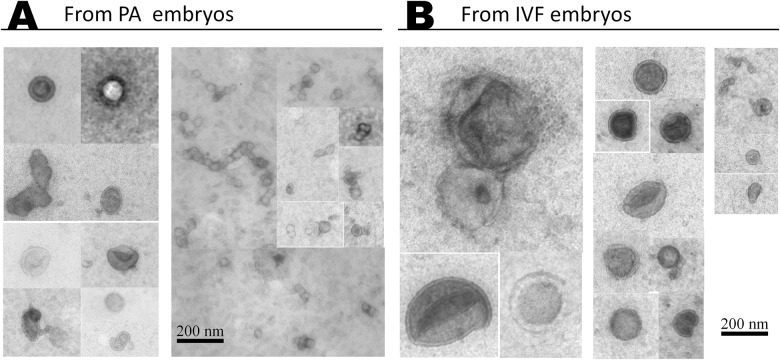
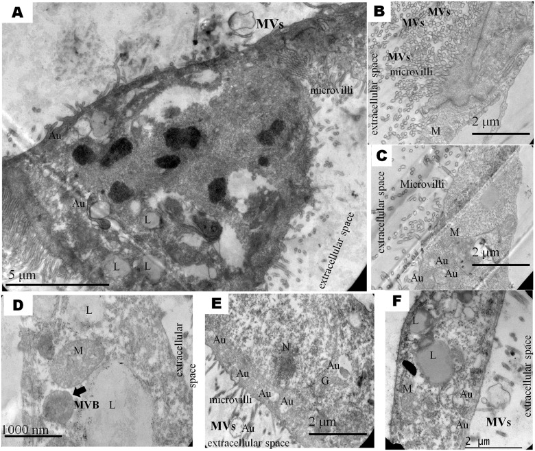
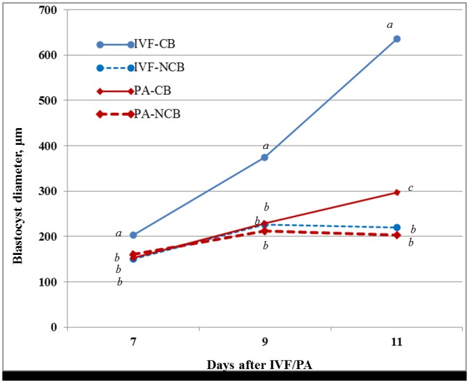
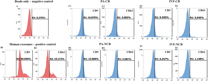
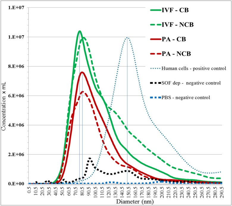
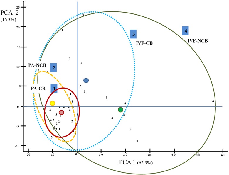
Extracellular vesicles (EVs) have been identified within different body fluids and cell culture media. However, there is very little information on the secretion of these vesicles during early embryonic development. The aims of this work were first to demonstrate the secretion of extracellular vesicles by pre-implantation bovine embryos and second to identify and characterize the population of EVs secreted by bovine blastocysts during the period from day seven to nine of embryo culture and its correlation with further embryo development up to day 11. Bovine embryos were produced by in vitro fertilization (IVF) or parthenogenetic activation (PA) and cultured until blastocyst stage. Blastocyst selection was performed at day 7 post IVF/PA considering two variables: stage of development and quality of embryos. Selected blastocysts were cultured in vitro for 48 hours in groups (exp. 1) or individually (exp. 2) in SOF media depleted of exosomes. At day 9 post IVF/PA the media was collected and EVs isolated by ultracentrifugation. Transmission electron microscopy revealed the presence of heterogeneous vesicles of different sizes and population: microvesicles (MVs) and exosomes (EXs) of rounded shape, enclosed by a lipid bi-layer and ranging from 30 to 385 nm of diameter. Flow cytometry analysis allowed identifying CD63 and CD9 proteins as exosome markers. Nanoparticle tracking analysis generated a large number of variables, which required the use of multivariate statistics. The results indicated that the concentration of vesicles is higher in those blastocysts with arrested development from day 9 up to day 11 of in vitro development (6.7 x 10⁸ particles/ml) derived from IVF (p <0.05), compared to PA blastocysts (4.7 x 10⁸ particles/ml). Likewise, the profile (concentration and diameter) of particles secreted by embryos derived from IVF were different from those secreted by PA embryos. In conclusion, we demonstrated that bovine blastocysts secrete MVs/EXs to the culture media. Data suggest that characteristics of the population of EVs vary depending on embryo competence.


Conflict of interest statement

Competing Interests: The authors have declared that no competing interests exist.

Figures

Fig 1
Fig 1. Morphology of EVs secreted by bovine blastocysts.
Representative pictures from transmission electron micrographs showing EVs secreted to culture media by IVF (B) and partenogenetic (A) derived blastocysts from day 7 to 9 of in vitro development.
Fig 2
Fig 2. Representative microphotograph showing ultrastructure of bovine IVF and PA derived blastocysts.
Nucleus (N; in E), mitochondria (M; in C, D, F), lipids (L; in A, D, F), autophagosome (Au; in A, C, E, F), multivesicular body MVB (arrow in D) and microvilli (in A, B, C, D, E) shown superficially of the cell.
Fig 3
Fig 3. Growth rate of blastocysts cultured in vitro according to their competence from day 7 to 11.
Values at the same day of embryo development carrying different superscripts are considered statistically significant at p < 0.05. CB: Competent blastocysts; NCB: non-competent blastocysts.
Fig 4
Fig 4. Flow cytometry analysis of EXs markers (CD9 and CD63).
Negative control-beads alone (A); positive control–EXs isolated from human embryonic kidney (HEK293) (B); EXs in competent (C and E) and non-competent (D and F) blastocysts.
Fig 5
Fig 5. Tracking of nanoparticles to determine size distribution of EVs populations from IVF and PA blastocysts, according to their competence.
The horizontal bars indicate the exosome size range. The EVs depleted SOF media and PBS used to resuspend isolated EVs, were included as negative controls. As a positive control, collected culture media from human cells was used.
Fig 6
Fig 6. Hierarchical Cluster Analysis (CA) of the first two principal component axes (Prin1 and Prin2) for EVs concentration according to vesicle size showing clusters based on blastocysts competence.
Each number represents the concentration of EVs from individual embryo from each experimental group: Number 1: Represents individual embryos from PA-CB; Number 2: Individual embryos from PA-NCB; Number 3: Individual embryos from IVF-CB; Number 4: Individual embryos from IVF-NCB.

Similar articles

Cited by

References

    1. van der Pol E, Böing AN, Harrison P, Sturk A, Nieuwland R. Classification, functions, and clinical relevance of extracellular vesicles. Pharmacol Rev. 2012;64(3): 676–705. 10.1124/pr.112.005983 - DOI - PubMed
    1. Tkach M, Théry C. Communication by extracellular vesicles: where we are and where we need to go. Cell. 2016;164(6): 1226–1232. 10.1016/j.cell.2016.01.043 - DOI - PubMed
    1. Yang Q, Diamond MP, Al-Hendy A. The emerging role of extracellular vesicle-derived miRNAs: Implication in cancer progression and stem cell related diseases. J Clin Epigenet. Articles in Press. 2016. 2(1): 13. - PMC - PubMed
    1. De Toro J, Herschlik L, Waldner C, Mongini C. Emerging roles of exosomes in normal and pathological conditions: new insights for diagnosis and therapeutic applications. Front Immunol. 2015;6: 203 10.3389/fimmu.2015.00203 - DOI - PMC - PubMed
    1. Dragovic RA, Gardiner C, Brooks AS, Tannetta DS, Ferguson DJ, Hole P, et al. Sizing and phenotyping of cellular vesicles using Nanoparticle Tracking Analysis. Nanomedicine. 2011;7(6): 780–788. 10.1016/j.nano.2011.04.003 - DOI - PMC - PubMed