. 2017 May 18;12(5):e0177879.
doi: 10.1371/journal.pone.0177879. eCollection 2017.

The PET and LIM1-2 domains of testin contribute to intramolecular and homodimeric interactions

Affiliations

The PET and LIM1-2 domains of testin contribute to intramolecular and homodimeric interactions

Stefano Sala et al. PLoS One. .

Abstract

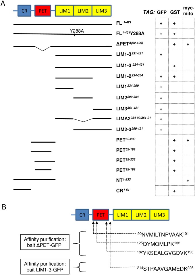
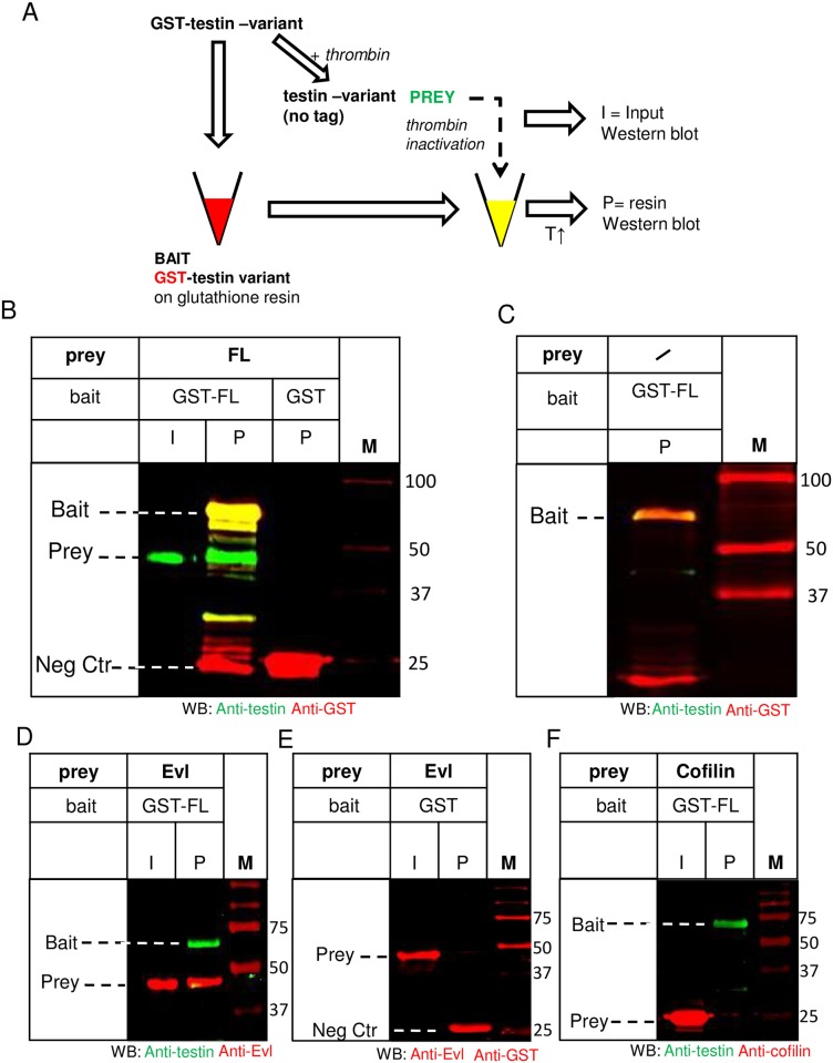
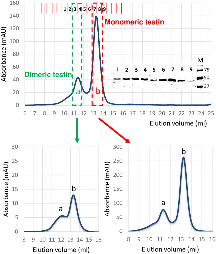
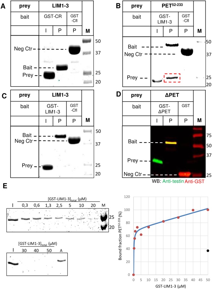
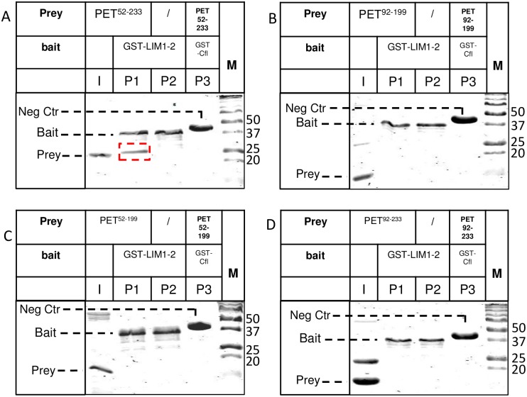
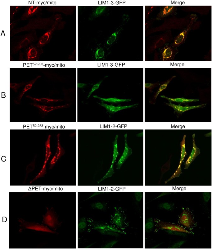
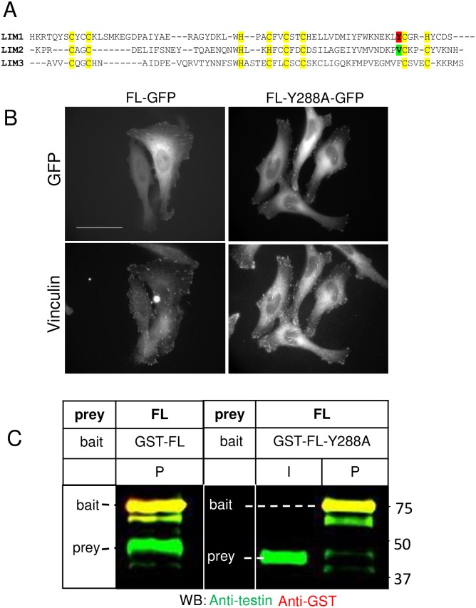
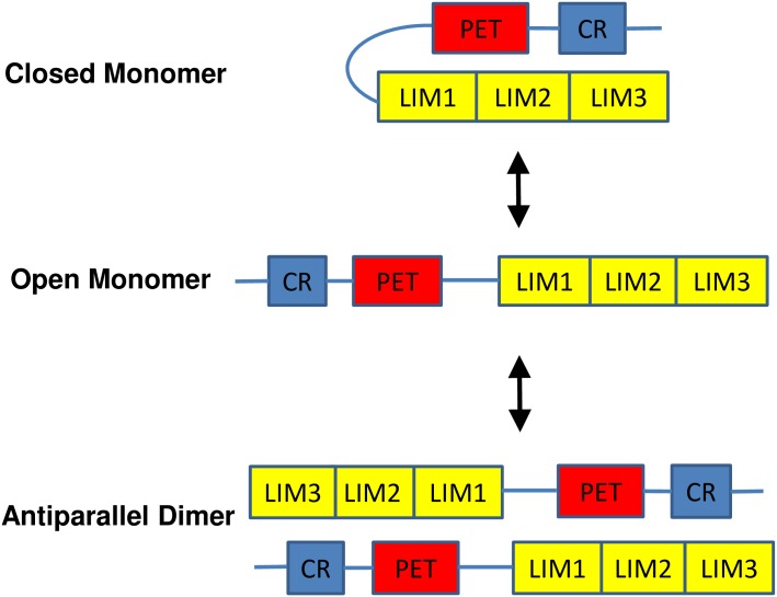
The focal adhesion protein testin is a modular scaffold and tumour suppressor that consists of an N-terminal cysteine rich (CR) domain, a PET domain of unknown function and three C-terminal LIM domains. Testin has been proposed to have an open and a closed conformation based on the observation that its N-terminal half and C-terminal half directly interact. Here we extend the testin conformational model by demonstrating that testin can also form an antiparallel homodimer. In support of this extended model we determined that the testin region (amino acids 52-233) harbouring the PET domain interacts with the C-terminal LIM1-2 domains in vitro and in cells, and assign a critical role to tyrosine 288 in this interaction.


Conflict of interest statement

Competing Interests: The authors have declared that no competing interests exist.

Figures

Fig 1
Fig 1. Tagged testin constructs.
A) Scheme of the structural domains of testin and of the different modular testin variants used in this study. Testin variants are coupled to GFP, GST or myc/mito (see Methods for details, ‘+’ indicates that a particular tagged version of the testin variant is used in one or more of the experiments presented in the manuscript). CR: cysteine rich; PET: Prickle, Espinas, Testin; LIM: Linl-1, Isl-1, Mec-3, NT: N-terminal part. Numbers indicate start and end of protein variants based on the numbering of full length (FL) human testin isoform 1 (Database ID: NP_056456.1). B) Sequence and location of testin peptides originating from endogenous testin that were identified using a mass spectrometry-based approach in the tryptic digests of complexes affinity purified using a GFP-nanobody from lysates of HeLa cells expressing ΔPET-GFP or LIM1-3-GFP as bait (PRIDE dataset identifier: PXD005058) [18,20].
Fig 2
Fig 2. Full length testin interacts with full length testin in vitro.
A) General Scheme of affinity purification used to produce data in several figures, to demonstrate an interaction of testin with other testin variants. In panel B of this figure, recombinant GST-FL was either used as bait on glutathione-sepharose or in parallel treated with thrombin to remove GST to be used as prey in an untagged form. Thrombin was inactivated prior to addition of this soluble form (input: I) to the resin with GST bound protein. After washing the resin, bound proteins were eluted with heated sample buffer and thus contain both bait and potential prey proteins (affinity purified: P). Proteins were detected either by Western Blotting (Figs 2B–2F and 4D) or Coomassie (Figs 4A–4C and 5A–5D), T = temperature. B) Immobilised recombinant GST-FL (bait) was incubated with soluble recombinant untagged FL (prey). A Western blot using anti-testin (green) and anti-GST (red) antibodies of the input (I) and proteins on the resin (P) is shown. FL prey (approx. 50 kDa) was present on the resin together with the bait GST-FL (lane ‘GST-FL/P’). Input (I) shows the untagged FL in solution. Recombinant immobilised GST was used as a negative control (lane ‘GST/P’). C) Western blot analysis (anti-testin (green), anti-GST (red)) of a mock buffer control incubated with immobilised GST-FL on resin. Similar as in B, the buffer contains inactivated thrombin but no soluble FL prey. Untagged FL is absent on the resin (P) (compare to Fig 2B, lane ‘GST-FL/P’) indicating that possible residual thrombin activity is not cleaving the GST-FL on the resin. D, E, F) Immobilised recombinant GST-FL (bait) was incubated with soluble untagged Evl (positive control, D) or cofilin (negative control, F) as preys. Immobilised GST was incubated with Evl (prey) and used as additional negative control (E). Western blot analysis of inputs (I) and proteins on the resin (P) is shown using anti testin (green), anti-Evl, anti-cofilin and anti-GST (red) antibodies. Untagged Evl (prey) is present on the GST-FL resin (lane P, 2D). Positions of bait, prey and negative control bait (Neg Ctr) are indicated in each panel. M: marker proteins (kDa).
Fig 3
Fig 3. Size exclusion chromatography reveals dimer formation of testin in vitro.
Top panel: Chromatogram of SEC of purified recombinant FL. Absorbance (280 nm) is plotted versus elution volume (ml). Two elution peaks are present (labelled a and b). Bold numbers 1–9 on top of chromatogram indicate the collected fractions which all contain testin as visualized by Coomassie staining after SDS-PAGE (see S1A Fig for column calibration). Bottom panels: Re-analysis by SEC of pooled fractions 3 and 4 (bottom left) or 7 and 8 (bottom right) of the primary SEC (boxed in top panel). Fractions 7 and 8 were first pooled and concentrated to a final concentration of 3,1 mg/ml prior to re-analysis with SEC. In both secondary runs two peaks eluted corresponding to dimeric (a) and monomeric (b) testin evidencing that both forms are in dynamic equilibrium with each other.
Fig 4
Fig 4. PET52-233 of testin directly interacts with LIM1-3 domains in vitro.
A-D) The experimental setup is similar as shown in Fig 2A. A recombinant GST-testin variant used as bait (GST-CR (A), GST-LIM1-3 (B, C) or GST-ΔPET (D)) was trapped on glutathione resin and presented in immobilised form to a second untagged testin-variant in solution used as prey (LIM1-3 (A, C), PET52-233 (B) or ΔPET (D)). Coomassie stained SDS-PAGE analysis (A-C) or western blot analysis (D) using anti-testin (green) and anti-GST (red) antibodies is shown. Input (I) shows the untagged prey protein prior to incubation with the resin. Lanes indicated with P show the proteins present on the resin (immobilized bait and/or bound prey) after the incubation. GST resin (D) or GST-cofilin resin (A-C) incubated with the same untagged testin variant as prey were used as negative controls. Untagged protein bound to the GST-testin variant immobilised on the resin is highlighted by a red box (B). Positions of bait, prey and negative control (Neg Ctr) bait are indicated in each panel, M: marker proteins (kDa). E) The indicated concentrations of GST-LIM1-3 immobilized on glutathione-sepharose beads were prepared and incubated with 2.5μM PET52-233. For each LIM1-3 concentration (indicated as [GST-LIM1-3]total (μM)), the level of unbound PET52-233 was analysed by Coomassie staining after SDS-PAGE (left). Aspecific binding was assessed by incubation of glutathione-sepharose beads, lacking LIM1-3, with a similar concentration of PET52-233 I: input, representing 2.5 μM PET52-233 in solution without incubation to LIM-1-3 coupled glutathione beads (reference for unbound 100% or bound 0%), A: aspecific binding of the PET52-233 ligand to beads without LIM1-3, M: molecular weight marker (kDa). (right) The % amounts of bound PET52-233 calculated from the measured intensities of the unbound material on gel (left) were plotted versus GST-LIM1-3 concentrations (graph right, red dots). The amount of aspecifically bound PET52-233 is represented by a black dot. The solid blue line in the graph is the fitted curve taking into account aspecific binding (see Materials and methods for details).
Fig 5
Fig 5. The PET domain of testin is not sufficient for interaction with LIM1-2 domains in vitro.
The experimental setup is similar as the scheme in Fig 2A. Immobilised GST-LIM1-2 on glutathione resin was incubated with untagged PET52-233(A), PET92-199(B), PET52-199 (C) or PET92-233 (D) in solution, used as preys. Coomassie stained SDS-PAGE analysis is shown: input (I) shows the untagged prey protein prior to incubation with the resin. Lanes indicated with P1 show the proteins present on the resin. We here included an extra negative control: the immobilised baits on the resin were mock-incubated with a solution lacking the soluble preys as in some cases the bait construct is prone to degradation during immobilization on the resin (lanes labelled P2). GST-cofilin resin was used as second negative control in each setup (lanes P3). Untagged prey protein PET52-233 bound to the GST- LIM1-2 testin variant immobilised on the resin is highlighted by a red box (A). Positions of bait, prey and negative control (Neg Ctr) bait are indicated in each panel, M: marker proteins (kDa).
Fig 6
Fig 6. The region 52–233 containing the PET domain of testin directly interacts with LIM1-2 domains in cells.
Immunofluorescence staining of myc/mito-coupled (bait) and GFP-coupled (prey) testin constructs in HeLa cells. Myc signal (red), GFP signal (green) and merge are shown for each condition. Colocalisation is observed for NT-myc/mito and LIM1-3-GFP (A), PET52-233-myc/mito and LIM1-3-GFP (B) and PET52-233-myc/mito and LIM1-2-GFP (C). Absence of colocalisation is observed for ΔPETΔ92-199-myc/mito and LIM1-2-GFP (D). See also S4 Fig.
Fig 7
Fig 7. Y288 is important for testin-testin interaction.
A) Multiple sequence alignment of the LIM domains of testin. Zinc coordinating cysteines (C) and histidines (H) are indicated with yellow highlight. Tyrosine (Y) 288 and valine (V) 348 are indicated in red and green respectively. This valine in LIM2 makes a hydrophobic stacking interaction with LIM3 based on the crystal structure in (Database id: 2xqn, [3]) B) Localization of GFP-coupled FL and FL-Y288A constructs in fixed HeLa cells. The GFP and vinculin signals are shown. FL and Y288A are distributed throughout the cytosol and present in focal adhesions. C) Immobilised recombinant GST -FL or GST-FL-Y288A (bait) was incubated with a similar amount of soluble recombinant untagged FL (prey) followed by western blot analysis of the protein on the resin using anti-testin (green) and anti-GST (red) antibodies. FL (approx. 50 kDa) is present on the resin (P) together with the immobilised GST-FL and immobilised GST-FL-Y288A. Input (I) shows the untagged FL prey in solution. Note the strongly reduced amount of bound FL when FL-Y288A mutant is used as bait. Positions of bait and prey are indicated in each panel. Molecular weights (kDa) are indicated.
Fig 8
Fig 8. Conformational model of the testin protein.
Testin can adopt an open active monomeric or a closed inactive monomeric conformation (as proposed by Garvalov et al. [1]) or an antiparallel dimeric conformation (this work). The activity status of the dimer is unknown (see Discussion). The interaction between PET52-233 and the LIM1-2 domains underlies formation of the dimer and/or closed monomer conformation.

References

    1. Garvalov BK, Higgins TE, Sutherland JD, Zettl M, Scaplehorn N, Köcher T, et al. The conformational state of Tes regulates its zyxin-dependent recruitment to focal adhesions. J Cell Biol. 2003;161(1):33–9. 10.1083/jcb.200211015 - DOI - PMC - PubMed
    1. Boëda B, Briggs DC, Higgins T, Garvalov BK, Fadden AJ, McDonald NQ, et al. Tes, a Specific Mena Interacting Partner, Breaks the Rules for EVH1 Binding. Mol Cell. 2007;28(6):1071–82. 10.1016/j.molcel.2007.10.033 - DOI - PubMed
    1. Boëda B, Knowles PP, Briggs DC, Murray-Rust J, Soriano E, Garvalov BK, et al. Molecular recognition of the Tes LIM2-3 domains by the actin-related protein Arp7A. J Biol Chem. 2011;286(13):11543–54. 10.1074/jbc.M110.171264 - DOI - PMC - PubMed
    1. Zheng Q, Zhao Y. The diverse biofunctions of LIM domain proteins: determined by subcellular localization and protein-protein interaction. Biol Cell. 2007;99(9):489–502. 10.1042/BC20060126 - DOI - PubMed
    1. Ma H, Weng D, Chen Y, Huang W, Pan K, Wang H, et al. Extensive analysis of D7S486 in primary gastric cancer supports TESTIN as a candidate tumor suppressor gene. Mol Cancer. 2010;9(1):190. - PMC - PubMed