. 2017 May 22;13(5):e1006398.
doi: 10.1371/journal.ppat.1006398. eCollection 2017 May.

FAS-associated factor-1 positively regulates type I interferon response to RNA virus infection by targeting NLRX1

Affiliations

FAS-associated factor-1 positively regulates type I interferon response to RNA virus infection by targeting NLRX1

Jae-Hoon Kim et al. PLoS Pathog. .

Erratum in

Abstract

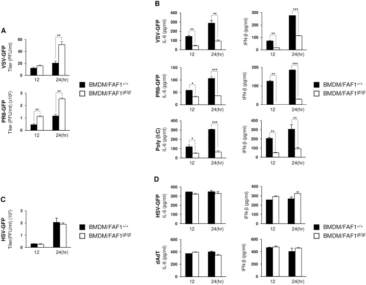
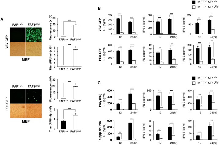
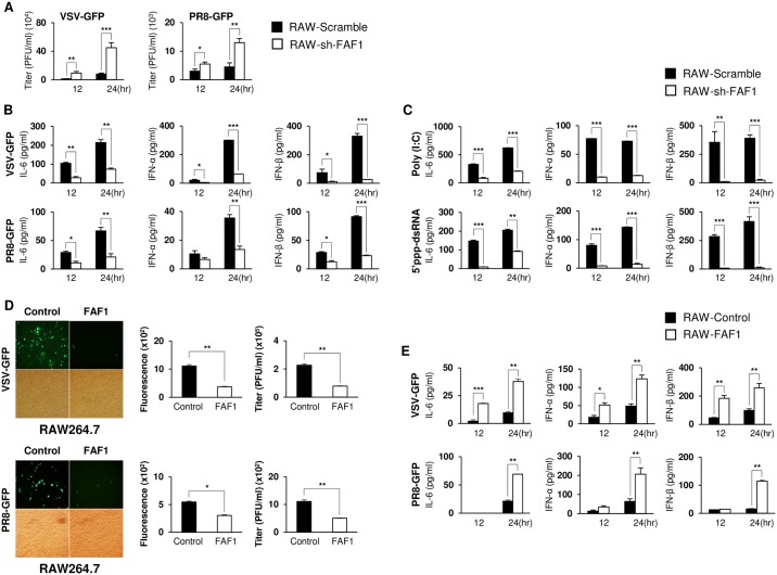
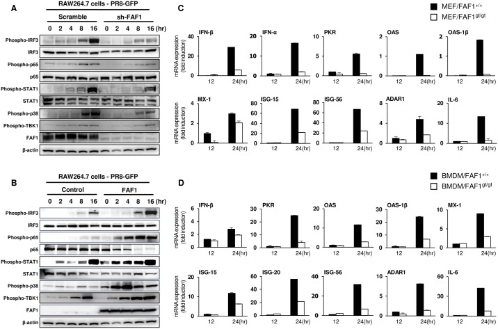
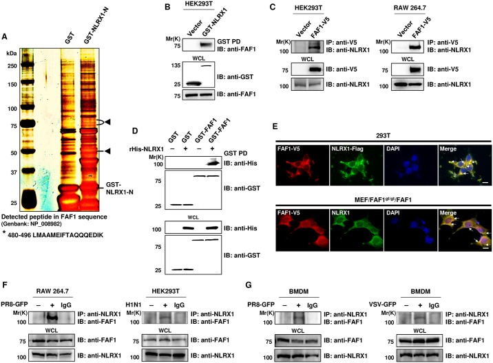
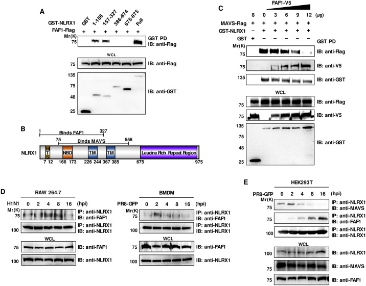
FAS-associated factor-1 (FAF1) is a component of the death-inducing signaling complex involved in Fas-mediated apoptosis. It regulates NF-κB activity, ubiquitination, and proteasomal degradation. Here, we found that FAF1 positively regulates the type I interferon pathway. FAF1gt/gt mice, which deficient in FAF1, and FAF1 knockdown immune cells were highly susceptible to RNA virus infection and showed low levels of inflammatory cytokines and type I interferon (IFN) production. FAF1 was bound competitively to NLRX1 and positively regulated type I IFN signaling by interfering with the interaction between NLRX1 and MAVS, thereby freeing MAVS to bind RIG-I, which switched on the MAVS-RIG-I-mediated antiviral signaling cascade. These results highlight a critical role of FAF1 in antiviral responses against RNA virus infection.


Conflict of interest statement

The authors have declared that no competing interests exist.

Figures

Fig 1
Fig 1. FAF1gt/gt mice are susceptible to virus infection and show suppressed immune responses.
(A) Wild-type mice (FAF1+/+) (n = 10) and FAF1 knockdown mice (FAF1gt/gt) (n = 10) were infected with VSV-Indiana (2 × 10⁸ pfu/mouse) via tail-vein injection and survival was monitored for 12 days. (B) Organs (spleen and brain) from FAF1+/+ (n = 10) and FAF1gt/gt (n = 10) mice were collected at 6 dpi with VSV-Indiana (2 × 10⁸ pfu/mouse) via tail-vein injection. Virus titers in supernatants of homogenized tissues were measured by plaque assay. (C) The viral load in supernatants of homogenized spleen, lung, liver, and brain tissues from FAF1+/+ (n = 4) and FAF1gt/gt (n = 4) mice infected with VSV-Indiana (2 × 10⁸ pfu/mouse) via tail-vein injection was measured by qRT-PCR at 6 dpi. (D and E) FAF1+/+ (n = 10) and FAF1gt/gt (n = 10) mice were infected with VSV-GFP (4 × 10⁸ pfu/mouse) via tail-vein injection. Sera were collected from the mice at indicated time points and the virus titer was determined by plaque assay. IFN- β and IL-6 were measured by ELISA. (F) PBMCs were isolated from whole peripheral blood of FAF1+/+ (PBMC/FAF1+/+; n = 5) and FAF1gt/gt (PBMC/FAF1gt/gt; n = 5) mice infected with VSV-GFP (4 × 10⁸ pfu/mouse) via tail-vein injection. Total RNA was extracted from PBMCs at 24 hpi and used for qRT-PCR analysis to determine levels of IFN-β, IFN-α, OAS, OAS-1β, MX-1, ISG-15, ISG-20, ISG-56, PML and GBP1 mRNA. All the mRNA expressions were normalized to GAPDH. Data are presented as the mean ± SEM. *p < 0.05, **p < 0.01, ***p < 0.001 (Student’s t test or log-rank test). Data are representative of at least two independent experiments.
Fig 2
Fig 2. FAF1 plays a role in antiviral activity in BMDMs upon RNA virus infection.
(A and B) Wild-type BMDMs (BMDM/FAF1+/+) or FAF1 knockdown BMDMs (BMDM/FAF1gt/gt) were stimulated with RNA virus (VSV-GFP (MOI = 2), PR8-GFP (MOI = 3)) or RNA stimulant (Poly (I:C) (20 μg/ml)). (C and D) BMDM/FAF1+/+ or BMDM/FAF1gt/gt were stimulated with DNA virus (HSV-GFP (MOI = 2)) or DNA stimulant (dAdT (1 μg/ml)). Virus titers (A and C) and IL-6 or IFN-β levels (B and D) were measured by plaque assay and ELISA, respectively. Data are presented as the mean ± SEM. *p < 0.05, **p < 0.01, ***p < 0.001 (Student’s t test). Data are representative of at least two independent experiments.
Fig 3
Fig 3. Knockdown of FAF1 augments viral replication and reduces Type I IFN secretion in MEFs.
(A) Wild-type MEFs (MEF/FAF1+/+) and FAF1 knockdown MEFs (MEF/FAF1gt/gt) were infected with VSV-GFP (MOI = 0.5) or PR8-GFP (MOI = 1). GFP expression of infected cells was visualized at 24 hpi, under a fluorescence microscopy (200 × magnification) and quantified using a fluorescence modulator. Virus titers were measured by plaque assay. (B and C) MEF/FAF1+/+ and MEF/FAF1gt/gt were infected with VSV-GFP (MOI = 0.5) or PR8-GFP (MOI = 1) (B) and treated with Poly (I:C) (20 μg/ml) or 5’ppp-dsRNA (1 μg/ml) (C). Levels of IL-6, IFN-α, and IFN-β in supernatants were measured by ELISA after 12 or 24 of infection or treatment. Data are presented as the mean ± SEM. *p < 0.05, **p < 0.01, ***p < 0.001 (Student’s t test). Data are representative of at least two independent experiments.
Fig 4
Fig 4. FAF1 plays a role in antiviral activity in RAW264.7 cells.
(A and B) RAW264.7 cells were infected with lentivirus harboring scramble and FAF1 shRNA to prepare control RAW264.7 (RAW-Scramble) and FAF1 knockdown RAW264.7 (RAW-sh-FAF1), respectively. Cells were infected with VSV-GFP (MOI = 2) or PR8-GFP (MOI = 3). After 12 and 24 hr, the virus titer was measured by plaque assay (A) and IL-6, IFN-α, and IFN-β levels in the supernatant were measured by ELISA (B). (C) Cells were treated with Poly (I:C) (20 μg/ml) or 5’ppp-dsRNA (1 μg/ml), and levels of IL-6, IFN-α, and IFN-β in the supernatant were measured by ELISA. (D and E) RAW264.7 cells were transfected with an empty IRES vector (control) and a FAF1-containing IRES plasmid. Stably expressing control (RAW-Control) and FAF1-overexpressing (RAW-FAF1) cells were infected with VSV-GFP (MOI = 1) or PR8-GFP (MOI = 2). At 24 hpi, GFP expression was visualized under a fluorescence microscopy (200 × magnification) and quantified using a fluorescence modulator. Virus titers were measured by plaque assay (D). Culture supernatants were collected at 12 h and 24 hpi, and IL-6, IFN-α, and IFN-β levels were measured by ELISA (E). Data are presented as the mean ± SEM. *p < 0.05, **p < 0.01, ***p < 0.001 (Student’s t test). Data are representative of at least two independent experiments.
Fig 5
Fig 5. FAF1 activates the Type I IFN signaling pathway and induces IFN-related gene expression.
(A and B) Control RAW264.7 (RAW-Scramble) and FAF1 knockdown RAW264.7 (RAW-sh-FAF1) cells (A) or control RAW264.7 (RAW-Control) and FAF1-overexpressing RAW264.7 (RAW-FAF1) cells (B) were infected with PR8-GFP (MOI = 2). At the indicated time points after infection, phosphorylated IRF3, p65, STAT1, p38 and TBK1, and total IRF3, p65 and STAT1 were measured in cell extracts by immunoblotting. β-actin was used to confirm equal loading of proteins. (C and D) Wild-type MEFs (MEF/FAF1+/+) and FAF1 knockdown MEFs (MEF/FAF1gt/gt) (C) and BMDMs isolated from FAF1+/+ (BMDM/FAF1+/+) and FAF1gt/gt (BMDM/FAF1gt/gt) mice (D) were infected with PR8-GFP (MOI = 1 and 3, respectively) for 12 hr, followed by total RNA extraction. Expression of mRNA encoding IFN-β, IFN-α, PKR, OAS, OAS-1β, MX-1, ISG-15, ISG-56, ADAR1 and IL-6 for MEFs and IFN-β, PKR, OAS, OAS-1β, MX-1, ISG-15, ISG-20, ISG-56, ADAR1 and IL-6 for BMDMs was analyzed by qRT-PCR. Data are presented as the mean ± SEM. Data are representative of at least two independent experiments.
Fig 6
Fig 6. FAF1 interacts with NLRX1.
(A) HEK293T cells were transfected with an empty GST vector (GST) or with the GST-NLRX1-N-terminal region (aa 1–225; GST-NLRX1-N). Proteins in the cell lysates were immunoprecipitated with GST beads and separated by 4–15% Nu-PAGE gels, followed by silver staining. Protein bands present exclusively in GST-NLRX1-N lane were excised from the gel and identified by mass spectrometry. The specific bands indicated by arrow heads were identified as FAF1. * indicates detected peptide corresponding to human FAF1 sequence. (B) WCL of HEK293T cells transfected with an empty GST vector or a GST-NLRX1 plasmid were subjected to a GST pull-down (GST PD) assay, followed by immunoblotting with anti-GST or anti-FAF1 antibodies. WCL were immunoblotted with anti-GST and anti-FAF1 antibodies. (C) WCL of HEK293T and RAW264.7 cells expressing FAF1-V5 proteins were immunoprecipitated with an anti-V5 antibody and immunoblotted with an anti-NLRX1 antibody. WCL were immunoblotted with anti-V5 and anti-NLRX1 antibodies. (D) In vitro binding assay, purified GST or GST-FAF1 protein immobilized on glutathione-conjugated Sepharose (GST) beads was incubated with the recombinant His tagged NLRX1 (rHis-NLRX1). After being pulled down, the bound proteins were subjected to immunoblotting with anti-GST and anti-His antibodies. WCL were immunoblotted with anti-GST and anti-His antibodies. (E) HEK293T cells transfected with FAF1-V5 and NLRX1-Flag plasmids were stained using anti-V5 and anti-Flag antibodies. FAF1-reconstituted MEFs (MEF/FAF1gt/gt/FAF1) were stained with anti-V5 and anti-NLRX1 antibodies. Secondary antibodies were labeled to identify localization of FAF1 (red; TRITC) and NLRX1 (green; FITC). Nuclei were stained using DAPI (blue). Yellow represents co-localization and marked in arrows. Bar, 10 μm. (F and G) RAW264.7, HEK293T and BMDMs cells were either mock-infected or infected with PR8-GFP (RAW264.7; MOI = 3, BMDMs; MOI = 3), H1N1 (HEK293T; MOI = 2) and VSV-GFP (BMDMs; MOI = 3MOI), and WCL were subjected to immunoprecipitation with an anti-NLRX1 antibody and control IgG, followed by immunoblot analysis with an anti-FAF1 antibody. WCL were immunoblotted with anti-FAF1 and anti-NLRX1 antibodies.
Fig 7
Fig 7. FAF1 inhibits the interaction between MAVS and NLRX1 after virus infection.
(A) HEK293T cells were transfected with the indicated GST-NLRX1 constructs (aa 1–156, 157-.327, 386–674, 675–975) and FAF1-Flag. GST pull-down (GST PD) was conducted followed by immunoblot analysis with an anti-Flag antibody. WCL were immunoblotted with anti-Flag and anti-GST antibodies. (B) Structure of NLRX1. The carton schematically indicates the positions of the mitochondrial targeting sequences (MTS, aa 7–12) nucleotide-binding domain (NBD, aa 166–173), transmembrane domain (TM, aa 226–244 and 367–385) and leucin-rich-repeat (LRR, aa 675–975) containing family member (known as NLR). FAF1 and MAVS bind to aa 1–327 and aa 75–556 region of NLRX1, respectively. (C) HEK293T cells were co-transfected with different doses of a FAF1-V5 plasmid and MAVS-Flag and GST-NLRX1 plasmids. NLRX1-binding proteins were detected by immunoprecipitation of NLRX1 by GST pull-down (GST PD), followed by immunoblot analysis with anti-FLAG, anti-V5, and anti-GST antibodies. WCL were immunoblotted with anti-V5, anti-Flag and anti-GST antibodies. (D) RAW264.7 and BMDM cells were infected with H1N1 (MOI = 2) and PR8-GFP (MOI = 3), respectively. Cells were harvested at the indicated time points and NLRX1 protein was immunoprecipitated with an anti-NLRX1, followed by immunoblot analysis with anti-FAF1 and anti-NLRX1 antibodies. WCL were immunoblotted with anti-FAF1 and anti-NLRX1 antibodies. (E) HEK293T cells were infected with PR8-GFP (MOI = 3) for the indicated time points. Infected cells were harvested and subjected to immunoprecipitation with an anti-NLRX1 antibody. Immunoprecipitates were then immunoblotted with anti-MAVS, anti-FAF1 and anti-NLRX1 antibodies. Expression of NLRX1, MAVS, and FAF1 in the WCL was confirmed using anti-NLRX1, anti-MAVS, and anti-FAF1 antibodies.

Similar articles

Cited by

References

    1. Ryu SW, Lee SJ, Park MY, Jun JI, Jung YK, et al. Fas-associated factor 1, FAF1, is a member of Fas death-inducing signaling complex. J Biol Chem. 2003; 278: 24003–24010. 10.1074/jbc.M302200200 - DOI - PubMed
    1. Song EJ, Yim SH, Kim E, Kim NS, Lee KJ. Human Fas-associated factor 1, interacting with ubiquitinated proteins and valosin-containing protein, is involved in the ubiquitin-proteasome pathway. Mol Cell Biol. 2005; 25: 2511–2524. 10.1128/MCB.25.6.2511-2524.2005 - DOI - PMC - PubMed
    1. Chu K, Niu X, Williams LT. A Fas-associated protein factor, FAF1, potentiates Fas-mediated apoptosis. Proc Natl Acad Sci U S A. 1995; 92: 11894–11898. - PMC - PubMed
    1. Park MY, Jang HD, Lee SY, Lee KJ, Kim E Fas-associated factor-1 inhibits nuclear factor-kappaB (NF-kappaB) activity by interfering with nuclear translocation of the RelA (p65) subunit of NF-kappaB. J Biol Chem. 2004; 279: 2544–2549. 10.1074/jbc.M304565200 - DOI - PubMed
    1. Park MY, Moon JH, Lee KS, Choi HI, Chung J, et al. FAF1 suppresses IkappaB kinase (IKK) activation by disrupting the IKK complex assembly. J Biol Chem. 2007; 282: 27572–27577. 10.1074/jbc.C700106200 - DOI - PubMed

MeSH terms