. 2017 May 25;12(5):e0178525.
doi: 10.1371/journal.pone.0178525. eCollection 2017.

The anti-inflammatory and antioxidant effects of melatonin on LPS-stimulated bovine mammary epithelial cells

Affiliations

The anti-inflammatory and antioxidant effects of melatonin on LPS-stimulated bovine mammary epithelial cells

Guang-Min Yu et al. PLoS One. .

Abstract

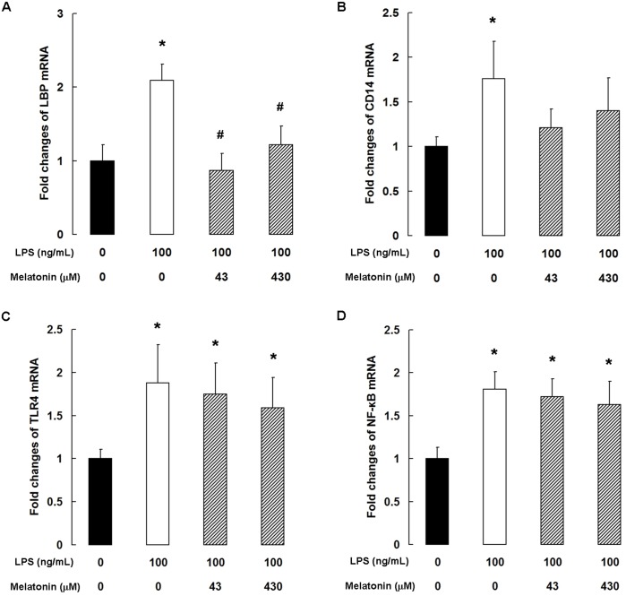
Mastitis is the most prevalent disease in dairy cattle worldwide and not only causes huge economic losses in the dairy industry but also threatens public health. To evaluate the therapeutic potential of melatonin in mastitis, we examined the ability of melatonin to protect bovine mammary epithelial cells (bMECs) from the harmful effects of lipopolysaccharide (LPS). We found that melatonin inhibited the LPS-binding protein-CD14-TLR4 signaling pathway in bMECs, which had opposing effects on pro-inflammatory and anti-inflammatory mediators. Melatonin decreased LPS-induced expression of pro-inflammatory cytokines, chemokines, and positive acute-phase proteins (APPs), including tumor necrosis factor-α, interleukin (IL)-1β, IL-6, granulocyte-monocyte colony-stimulating factor, chemokine CC motif ligand (CCL)2, CCL5, serum amyloid A, haptoglobin, C-reactive protein, ceruloplasmin, and α-1 antitrypsin, and increased expression of the anti-inflammatory cytokine IL-1Ra and the negative APP fibrinogen. In addition, melatonin increased dityrosine levels but suppressed nitrite levels by upregulating the expression of Nrf2 and heme oxygenase-1 in the Nrf2 antioxidant defense pathway. Finally, melatonin administration increased the viability of LPS-stimulated bMECs. These results suggest that melatonin protects bMECs from LPS-induced inflammatory and oxidant stress damage and provide evidence that melatonin might have therapeutic utility in mastitis.


Conflict of interest statement

Competing Interests: The authors have declared that no competing interests exist.

Figures

Fig 1
Fig 1. Melatonin prevents LPS-induced death of cultured bMECs.
(A) Fluorescence microscopic images of cell double-staining. Data are representative of three independent experiments. Scale bar: 20 μm. (B) cell viability analyzed by MTT assay. Data are the mean ± SD of three independent experiments, each performed in sextuplicate. qPCR analysis of (C) Bcl-2 and (D) Bax. Data are the mean ± SD of five independent experiments, each performed in duplicate. *P < 0.05 vs the control group; #P < 0.05 vs the 100 ng/mL LPS group; $P < 0.05 vs the 100 ng/mL LPS + 43 μM melatonin group.
Fig 2
Fig 2. Melatonin inhibits the TLR4 signaling pathway in LPS-stimulated bMECs.
qPCR analysis of (A) TLR4, (B) LBP, (C) CD14, and (D) NF-κB. Data are the mean ± SD of five independent experiments, each performed in duplicate. *P < 0.05 vs the control group; #P < 0.05 vs the 100 ng/mL LPS group.
Fig 3
Fig 3. Melatonin modulates LPS-induced cytokine expression.
qPCR analysis of (A) TNF-α, (B) IL-1β, (C) IL-6, (D) IL-8, (E) GM-CSF, and (F) IL-1Ra. Data are the mean ± SD of five independent experiments, each performed in duplicate. *P < 0.05 vs the control group; #P < 0.05 vs the 100 ng/mL LPS group; $P < 0.05 vs the 100 ng/mL LPS + 43 μM melatonin group.
Fig 4
Fig 4. Melatonin inhibits LPS-induced chemokine expression.
qPCR analysis of (A) CCL2, (B) CCL3, (C) CCL5, (D) CCL20, (E) CXCL1, and (F) CXCL2. Data are the mean ± SD of five independent experiments, each performed in duplicate. *P < 0.05 vs the control group; #P < 0.05 vs the 100 ng/mL LPS group; $P < 0.05 vs the 100 ng/mL LPS + 43 μM melatonin group.
Fig 5
Fig 5. Melatonin modulates LPS-induced acute-phase protein expression.
qPCR analysis of (A) SAA, (B) haptoglobin, (C) ceruloplasmin, (D) CRP, (E) α-1 antitrypsin, and (F) fibrinogen. Data are the mean ± SD of five independent experiments, each performed in duplicate. *P < 0.05 vs the control group; #P < 0.05 vs the 100 ng/mL LPS group; $P < 0.05 vs the 100 ng/mL LPS + 43 μM melatonin group.
Fig 6
Fig 6. Melatonin reduces oxidative stress in LPS-stimulated bMECs.
qPCR analysis of (A) Nrf2, (B) HO-1, and (C) iNOS. Data are the mean ± SD of five independent experiments, each performed in duplicate. (D) dityrosine level and (E) nitrite level in the culture medium. Data are the mean ± SD of six independent experiments, each performed in duplicate. *P < 0.05 vs the control group; #P < 0.05 vs the 100 ng/mL LPS group; $P < 0.05 vs the 100 ng/mL LPS + 43 μM melatonin group.

Similar articles

Cited by

References

    1. Tothova C, Nagy O, Kovac G. Acute phase proteins and their use in the diagnosis of diseases in ruminants: a review. Vet. Med.-Czech 2014;59:163–180.
    1. Hogeveen H, Huijps K, Lam TJ. Economic aspects of mastitis: new developments. N. Z. Vet. J. 2011;59:16–23. 10.1080/00480169.2011.547165 - DOI - PubMed
    1. Ellah MRA. Role of free radicals and antioxidants in mastitis. J. Adv. Vet. Res. 2013;3:1–7.
    1. Osman KM, Hassan HM, Ibrahim IM, Mikhail MMS. The impact of staphylococcal mastitis on the level of milk IL-6, lysozyme and nitric oxide. Comp. Immunol. Microb. 2010;33:85–93. - PubMed
    1. Wellnitz O, Reith P, Haas SC, Meyer HHD. Immune relevant gene expression of mammary epithelial cells and their influence on leukocyte chemotaxis in response to different mastitis pathogens. Vet. Med.-Czech 2006;51:125–132.

MeSH terms