. 2017 May 19;13(5):e1006402.
doi: 10.1371/journal.ppat.1006402. eCollection 2017 May.

Seminal plasma induces inflammation and enhances HIV-1 replication in human cervical tissue explants

Affiliations

Seminal plasma induces inflammation and enhances HIV-1 replication in human cervical tissue explants

Andrea Introini et al. PLoS Pathog. .

Erratum in

Abstract

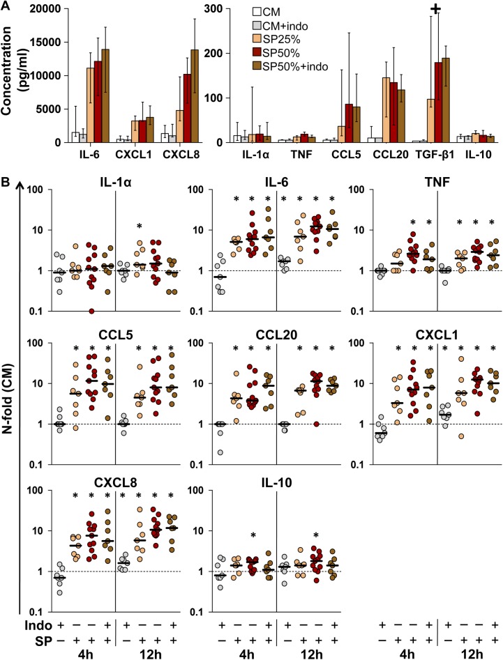
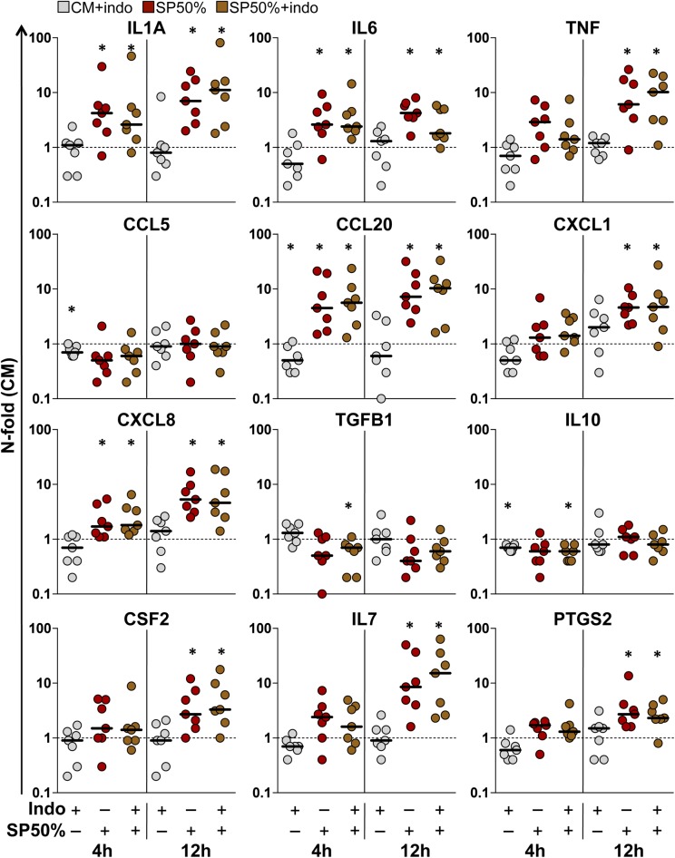
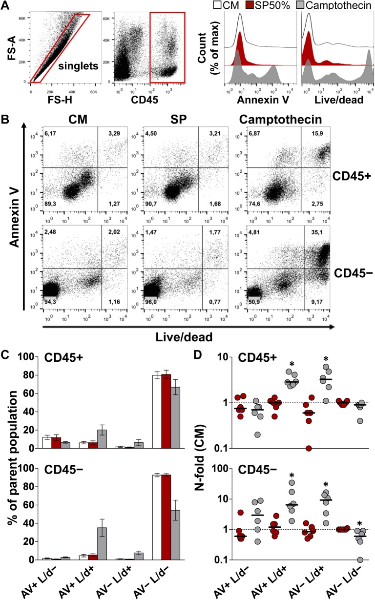
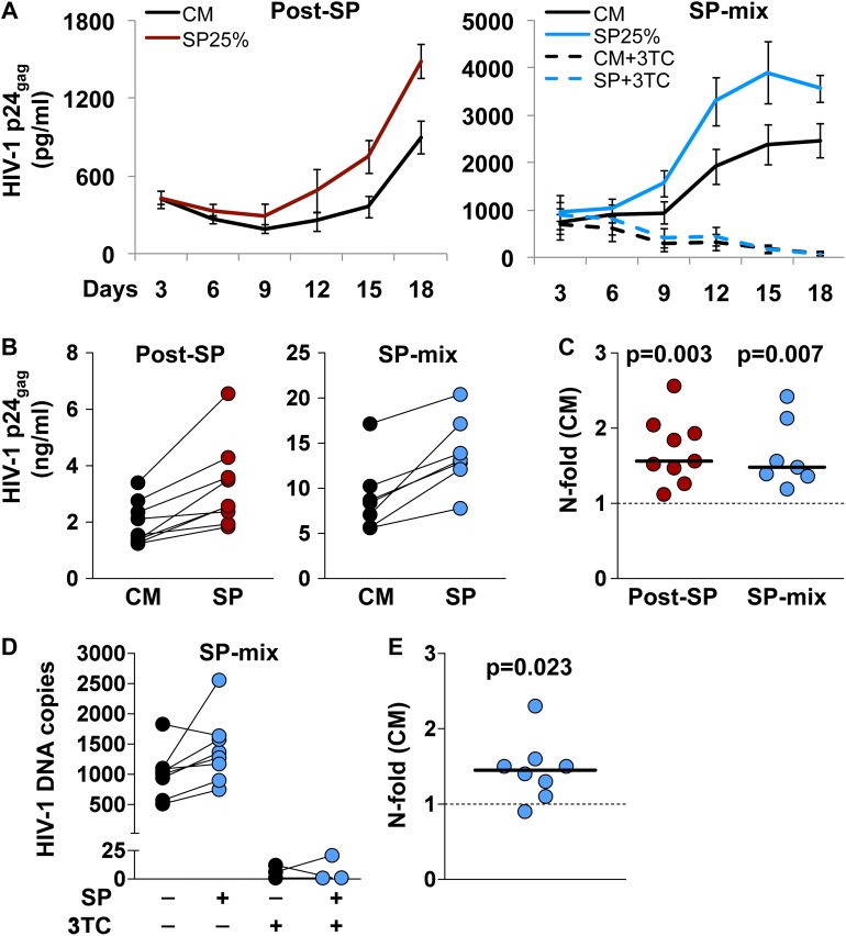
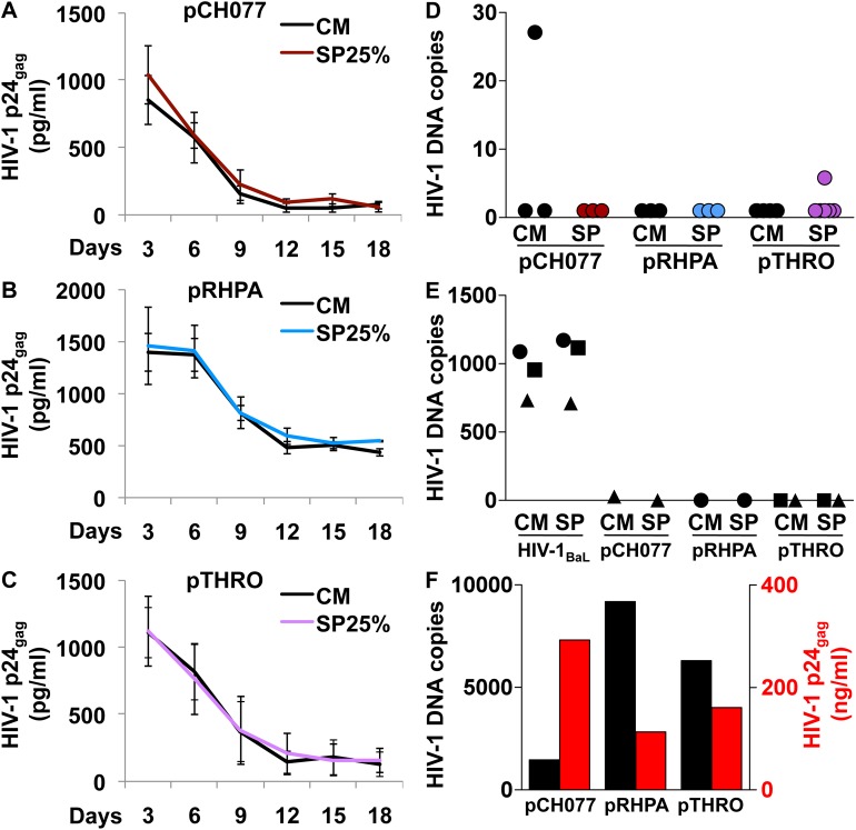
The most immediate and evident effect of mucosal exposure to semen in vivo is a local release of proinflammatory mediators accompanied by an influx of leukocytes into the female genital mucosa (FGM). The implication of such response in HIV-1 transmission has never been addressed due to limitations of currently available experimental models. Using human tissue explants from the uterine cervix, we developed a system of mucosal exposure to seminal plasma (SP) that supports HIV-1 replication. Treatment of ectocervical explants with SP resulted in the upregulation of inflammatory and growth factors, including IL-6, TNF, CCL5, CCL20, CXCL1, and CXCL8, and IL1A, CSF2, IL7, PTGS2, as evaluated by measuring protein levels in explant conditioned medium (ECM) and gene expression in tissue. SP treatment was also associated with increased recruitment of monocytes and neutrophils, as observed upon incubation of peripheral blood leukocytes with ECM in a transwell system. To evaluate the impact of the SP-mediated response on local susceptibility to HIV-1, we infected ectocervical explants with the CCR5-tropic variant HIV-1BaL either in the presence of SP, or after explant pre-incubation with SP. In both experimental settings SP enhanced virus replication as evaluated by HIV-1 p24gag released in explant culture medium over time, as well as by HIV-1 DNA quantification in explants infected in the presence of SP. These results suggest that a sustained inflammatory response elicited by SP soon after coitus may promote HIV-1 transmission to the FGM. Nevertheless, ectocervical tissue explants did not support the replication of transmitted/founder HIV-1 molecular clones, regardless of SP treatment. Our system offers experimental and analytical advantages over traditional models of HIV-1 transmission for the study of SP immunoregulatory effect on the FGM, and may provide a useful platform to ultimately identify new determinants of HIV-1 infection at this site.


Conflict of interest statement

The authors have declared that no competing interests exist.

Figures

Fig 1
Fig 1. Cytokine concentration in ectocervical tissue explant conditioned medium (ECM).
A) Cytokine concentration (pg/ml) was measured in ECM of ectocervical explants incubated with culture medium (CM), seminal plasma (SP) 25% or SP50% in the presence or absence of indomethacin (indo) 10μM for 4 h, followed by an additional 12 h-incubation with medium only. Bars represent median with interquartile range (IQR). + excluded from the analysis (TGF-β1). B) N-fold change in ECM cytokine concentration of explants treated with SP and/or indomethacin for 4 or 12 h, compared to donor-matched explants incubated with CM. Bars indicate median values. Asterisks denote a statistically significant difference with CM (Wilcoxon signed rank test, p<0.05). CM (white, n = 11), CM+indomethacin (gray, n = 7), SP25% (orange, n = 7), SP50% (red, n = 11), and SP50%+indomethacin (brown, n = 7).
Fig 2
Fig 2. Gene expression in ectocervical tissue explants.
N-fold change in gene expression measured in ectocervical explants incubated with indomethacin (indo) 10μM (gray), and seminal plasma (SP) 50% without (red) or with (brown) indomethacin for 4 or 12 h, compared to donor-matched explants incubated with culture medium (CM) (n = 7), followed by an additional 12 h-incubation with medium only. Bars indicate median values. Asterisks denote a significant difference with CM (Wilcoxon signed rank test, p<0.05).
Fig 3
Fig 3. Transwell migration of peripheral blood leukocytes (PBL).
Mononuclear cells and granulocytes were isolated from blood, pooled (i.e. PBL) and incubated in a transwell system for 2 h with explant conditioned medium (ECM) from donor-matched ectocervical explants incubated with culture medium (CM-ECM) or seminal plasma (SP-ECM). Transmigrated cells were immunophenotyped and enumerated by flow cytometry (see S5 Fig). A) Fraction of transmigrated cells out of total number of PBL loaded into a transwell insert for each analyzed cell population (input). PBL were incubated with CM-ECM (white), SP-ECM (red), and with medium only (grey) and medium supplemented with FBS 10% (yellow) as negative and positive controls respectively. Bars represent mean with s.e.m (n = 6). B) N-fold change in the fraction of transmigrated PBL incubated with SP-ECM compared to that of donor-matched CM-ECM. Bars indicate median values. p<0.05 denotes a significant difference with CM (Wilcoxon signed rank test). C) Expression levels of the chemokine receptor CCR5 on transmigrated monocytes. Top, peaks represent PBL untreated cultured (ctrl, gray), cultured with CM-ECM (CM, white), cultured with SP-ECM (SP, red), and unstained control (black) from one representative experiment. Middle, CCR5 mean fluorescence intensity (MFI). Bars indicate median values. Bottom, n-fold change in CCR5 MFI on PBL cultured with ECM compared to untreated cultured PBL (ctrl). Lines connect measurements obtained from donor-matched ECM. p<0.05 denotes a significant difference with ctrl (Wilcoxon signed rank test).
Fig 4
Fig 4. Viability of cells isolated from ectocervical tissue explants.
Cells were isolated from ectocervical explants after incubation with culture medium (CM, white) or seminal plasma (SP, red) 50% for 12 h, followed by an additional 12 h-incubation with medium only. As a positive control, camptothecin 100 μM (grey) was used to induce apoptosis upon explant treatment for 24 h. Cells were stained with annexin V (AV) and an amine-reactive dye (Live/dead, L/d) and analyzed by flow cytometry. A) Representative dot plots of the gating strategy. Expression analysis of the two selected cell death markers was conducted on events phenotyped as immune (CD45+) and non-immune (CD45-) cells. Histograms depict the expression level of the cell death markers on singlets, which comprise both CD45+ and CD45- cells, isolated from donor-matched explants of one representative experiment. B) Dot plots of the fraction of cells expressing the two analyzed cell death markers among CD45+ (upper panel) and CD45- (lower panel) cells isolated from donor-matched explants of one representative experiment. C) Fraction of CD45+ (upper chart) and CD45- (lower chart) cells expressing different combinations of the two analyzed cell death markers. Bars represent mean with s.e.m (n = 6). D) N-fold change in the fraction of CD45+ (upper chart) and CD45- (lower chart) cells expressing different combinations of the two analyzed cell death markers from explants treated with SP (red) and camptothecin (grey) compared to untreated donor-matched explants (CM). Bars indicate median values. Asterisks denote a statistically significant difference with CM (Wilcoxon signed rank test, p<0.05).
Fig 5
Fig 5. Infection of ectocervical tissue explants with HIV-1BaL.
Infection of ectocervical explants with HIV-1BaL was independently performed after seminal plasma (SP) 25% treatment (post-SP,red), or in the presence of SP25% (SP-mix, blue) (see S1B Fig). In selected experiments, explants infected in the presence of SP were treated with lamivudine (3TC) 10μM throughout culture time. A) Kinetics of HIV-1BaL replication in explants treated with SP (colored line) or culture medium (CM) (black line). Virus replication was evaluated as p24gag concentration in explant culture medium over 18 days. Represented are mean values with s.e.m. (n = 9 for post-SP; n = 8 for SP-mix; n = 3 for SP-mix+3TC). B) Cumulative p24gag production over culture time. Lines connect measurements obtained from donor-matched explants. C) N-fold change in cumulative p24gag production in SP-treated explants compared to donor-matched untreated explants. Bars indicate median values. p<0.05 denotes a significant difference with CM (Wilcoxon signed rank test). D) HIV-1 DNA quantification in explants infected with a mix of HIV and SP or CM, cultured in the presence or the absence of 3TC, and harvested at the end of culture (day18). HIV-1 DNA copy numbers were normalized to the amount of the single-copy gene HBB. Lines connect measurements obtained from donor-matched explants. E) N-fold change in HIV-1 DNA copy numbers in SP-treated explants compared to donor-matched untreated explants. The bar indicates median value. p<0.05 denotes a significant difference with CM (Wilcoxon signed rank test).
Fig 6
Fig 6. Infection of ectocervical tissue explants with transmitted/founder (T/F) HIV-1.
Infection of ectocervical explants with pCH077.t/2627 (pCH077), pRHPA.c/2635 (pRHPA), and pTHRO.c/2626 (pTHRO) was performed after an initial treatment with seminal plasma (SP) 25% (see S1B Fig). A-C) Kinetics of virus replication in explants treated with SP (colored line) or culture medium (CM) (black line). Virus replication was evaluated as p24gag concentration in explant culture medium over 18 days. Represented are mean values with s.e.m. (n = 3 for pCH077; n = 3 for pRHPA; n = 4 for pTHRO). D-E) HIV-1 DNA copy numbers in infected explants harvested at the end of culture (day18). Values were normalized to the amount of the single-copy gene HBB. E) Donor-matched explants were independently infected with HIV-1BaL and at least one T/F virus (n = 3). Donor-matched explants are indicated with the same symbol. F) Exogenously activated PBMCs were infected with T/F HIV-1 produced in 293T cells. HIV-1 replication was evaluated as p24gag concentration in culture supernatant (red) and HIV-1 DNA copy numbers (black) in cells harvested at the end of culture (day 8–10 post-infection) (n = 1).

Similar articles

Cited by

References

    1. Hladik F, McElrath MJ (2008) Setting the stage: host invasion by HIV. Nat Rev Immunol 8: 447–457. doi: 10.1038/nri2302 - DOI - PMC - PubMed
    1. Roan NR, Munch J (2015) Improving preclinical models of HIV microbicide efficacy. Trends Microbiol 23: 445–447. doi: 10.1016/j.tim.2015.05.001 - DOI - PMC - PubMed
    1. Southern PJ (2013) Missing out on the biology of heterosexual HIV-1 transmission. Trends Microbiol 21: 245–252. doi: 10.1016/j.tim.2013.02.002 - DOI - PubMed
    1. Vanpouille C, Arakelyan A, Margolis L (2012) Microbicides: still a long road to success. Trends Microbiol 20: 369–375. doi: 10.1016/j.tim.2012.05.005 - DOI - PMC - PubMed
    1. Doncel GF, Anderson S, Zalenskaya I (2014) Role of semen in modulating the female genital tract microenvironment-implications for HIV transmission. Am J Reprod Immunol 71: 564–574. doi: 10.1111/aji.12231 - DOI - PubMed

MeSH terms