

Gallium nanoparticles facilitate phagosome maturation and inhibit growth of virulent Mycobacterium tuberculosis in macrophages
- PMID: 28542623
- PMCID: PMC5436895
- DOI: 10.1371/journal.pone.0177987
Gallium nanoparticles facilitate phagosome maturation and inhibit growth of virulent Mycobacterium tuberculosis in macrophages
Abstract
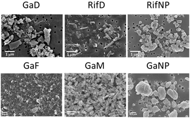
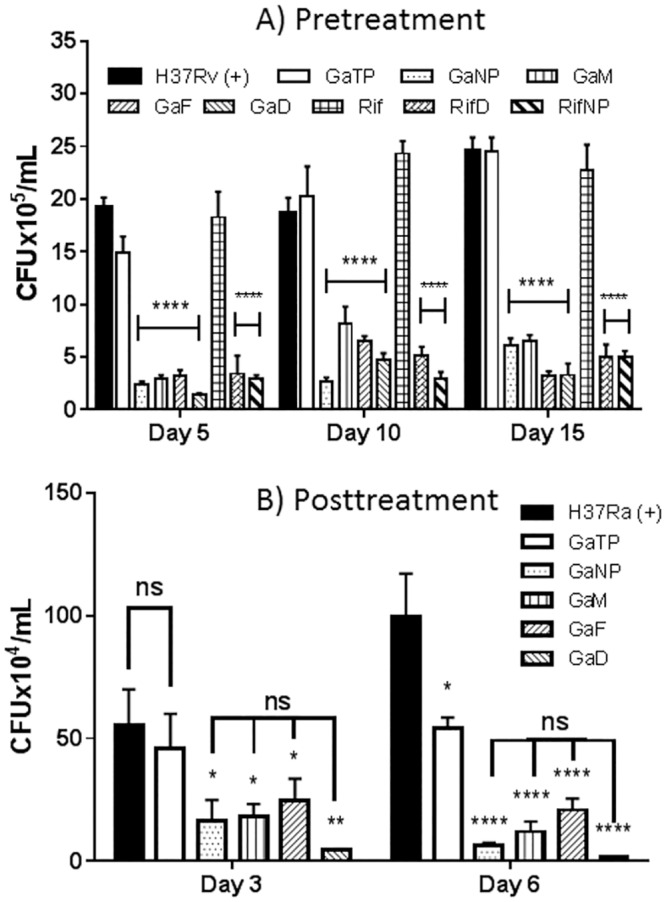
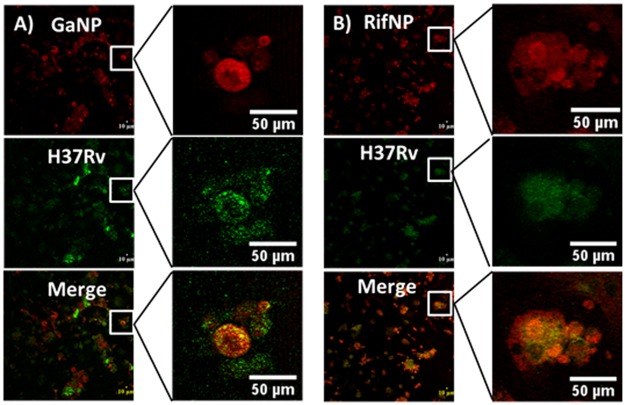
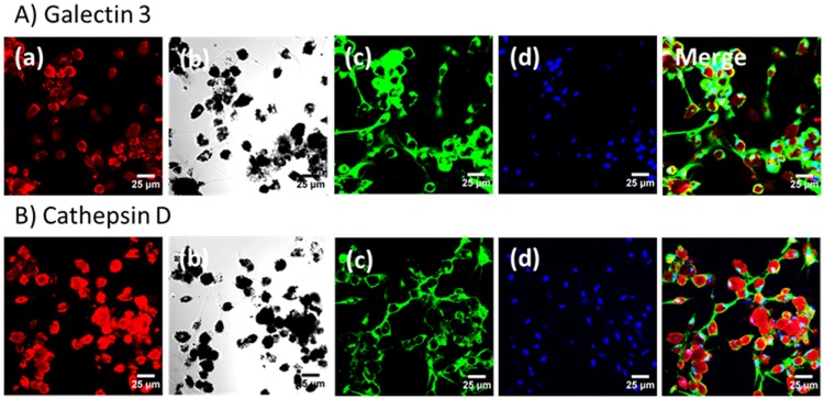
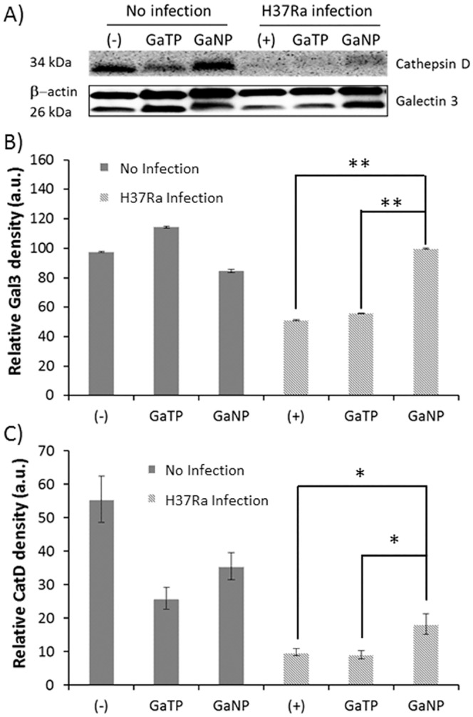
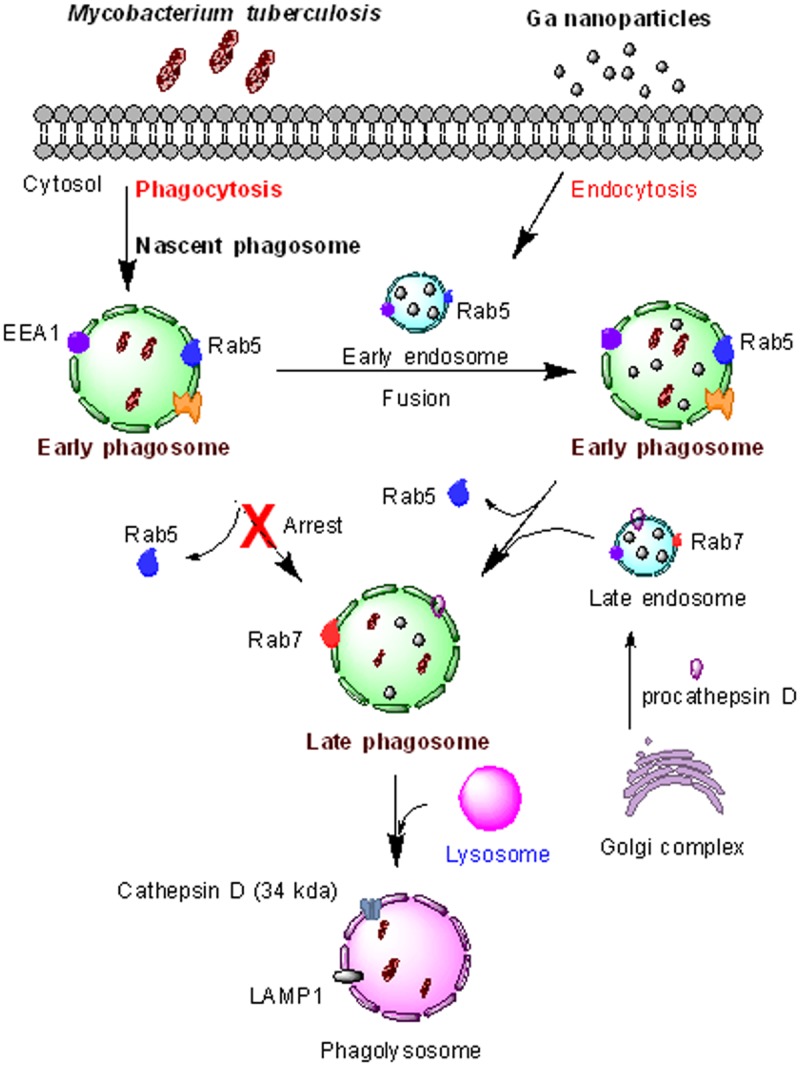
New treatments and novel drugs are required to counter the growing problem of drug-resistant strains of Mycobacterium tuberculosis (M.tb). Our approach against drug resistant M.tb, as well as other intracellular pathogens, is by targeted drug delivery using nanoformulations of drugs already in use, as well as drugs in development. Among the latter are gallium (III) (Ga)-based compounds. In the current work, six different types of Ga and rifampin nanoparticles were prepared in such a way as to enhance targeting of M.tb infected-macrophages. They were then tested for their ability to inhibit growth of a fully pathogenic strain (H37Rv) or a non-pathogenic strain (H37Ra) of M.tb. Encapsulating Ga in folate- or mannose-conjugated block copolymers provided sustained Ga release for 15 days and significantly inhibited M.tb growth in human monocyte-derived macrophages. Nanoformulations with dendrimers encapsulating Ga or rifampin also showed promising anti-tuberculous activity. The nanoparticles co-localized with M.tb containing phagosomes, as measured by detection of mature cathepsin D (34 kDa, lysosomal hydrogenase). They also promoted maturation of the phagosome, which would be expected to increase macrophage-mediated killing of the organism. Delivery of Ga or rifampin in the form of nanoparticles to macrophages offers a promising approach for the development of new therapeutic anti-tuberculous drugs.
Conflict of interest statement
Figures


Similar articles
-
Treatment of Virulent Mycobacterium tuberculosis and HIV Coinfected Macrophages with Gallium Nanoparticles Inhibits Pathogen Growth and Modulates Macrophage Cytokine Production.mSphere. 2019 Jul 24;4(4):e00443-19. doi: 10.1128/mSphere.00443-19. mSphere. 2019. PMID: 31341073 Free PMC article.
-
Ga(III) Nanoparticles Inhibit Growth of both Mycobacterium tuberculosis and HIV and Release of Interleukin-6 (IL-6) and IL-8 in Coinfected Macrophages.Antimicrob Agents Chemother. 2017 Mar 24;61(4):e02505-16. doi: 10.1128/AAC.02505-16. Print 2017 Apr. Antimicrob Agents Chemother. 2017. PMID: 28167548 Free PMC article.
-
Prolonged-acting, multi-targeting gallium nanoparticles potently inhibit growth of both HIV and mycobacteria in co-infected human macrophages.Sci Rep. 2015 Mar 6;5:8824. doi: 10.1038/srep08824. Sci Rep. 2015. PMID: 25744727 Free PMC article.
-
[Development of antituberculous drugs: current status and future prospects].Kekkaku. 2006 Dec;81(12):753-74. Kekkaku. 2006. PMID: 17240921 Review. Japanese.
-
Mycobacteria, metals, and the macrophage.Immunol Rev. 2015 Mar;264(1):249-63. doi: 10.1111/imr.12265. Immunol Rev. 2015. PMID: 25703564 Free PMC article. Review.
Cited by
-
Brief update on endocytosis of nanomedicines.Adv Drug Deliv Rev. 2019 Apr;144:90-111. doi: 10.1016/j.addr.2019.08.004. Epub 2019 Aug 13. Adv Drug Deliv Rev. 2019. PMID: 31419450 Free PMC article. Review.
-
Advancement of Gallium and Gallium-Based Compounds as Antimicrobial Agents.Front Bioeng Biotechnol. 2022 Feb 4;10:827960. doi: 10.3389/fbioe.2022.827960. eCollection 2022. Front Bioeng Biotechnol. 2022. PMID: 35186906 Free PMC article. Review.
-
Iron/Heme Metabolism-Targeted Gallium(III) Nanoparticles Are Active against Extracellular and Intracellular Pseudomonas aeruginosa and Acinetobacter baumannii.Antimicrob Agents Chemother. 2019 Mar 27;63(4):e02643-18. doi: 10.1128/AAC.02643-18. Print 2019 Apr. Antimicrob Agents Chemother. 2019. PMID: 30782994 Free PMC article.
-
In Vitro Efficacy of Free and Nanoparticle Formulations of Gallium(III) meso-Tetraphenylporphyrine against Mycobacterium avium and Mycobacterium abscessus and Gallium Biodistribution in Mice.Mol Pharm. 2018 Mar 5;15(3):1215-1225. doi: 10.1021/acs.molpharmaceut.7b01036. Epub 2018 Feb 16. Mol Pharm. 2018. PMID: 29421865 Free PMC article.
-
Efficacy and Possible Mechanism(s) of Action of Gallium Tetraphenylporphyrin Nanoparticles against HIV-TB Coinfection in an In Vitro Granuloma Structure Model.ACS Infect Dis. 2024 Dec 13;10(12):4279-4290. doi: 10.1021/acsinfecdis.4c00639. Epub 2024 Nov 5. ACS Infect Dis. 2024. PMID: 39499869 Free PMC article.
References
-
- WHO. Global Tuberculosis Report. World Health Organization. 2015; Geneva:Switzerland.
-
- Alexander PE, De P. The emergence of extensively drug-resistant tuberculosis (TB): TB/HIV coinfection, multidrug-resistant TB and the resulting public health threat from extensively drug-resistant TB, globally and in Canada. The Canadian journal of infectious diseases & medical microbiology = Journal canadien des maladies infectieuses et de la microbiologie medicale / AMMI Canada. 2007;18(5):289–91. ; - PMC - PubMed
MeSH terms
Substances
Grants and funding
LinkOut - more resources
Full Text Sources
Other Literature Sources
