. 2017 May 19;13(5):e1006385.
doi: 10.1371/journal.ppat.1006385. eCollection 2017 May.

Platelet proteome reveals novel pathways of platelet activation and platelet-mediated immunoregulation in dengue

Affiliations

Platelet proteome reveals novel pathways of platelet activation and platelet-mediated immunoregulation in dengue

Monique Ramos de Oliveira Trugilho et al. PLoS Pathog. .

Abstract

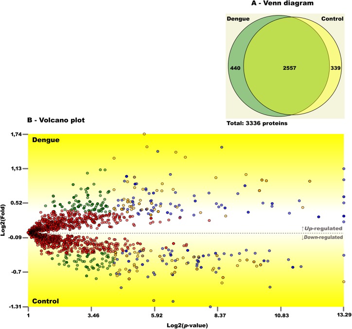
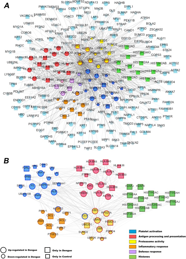
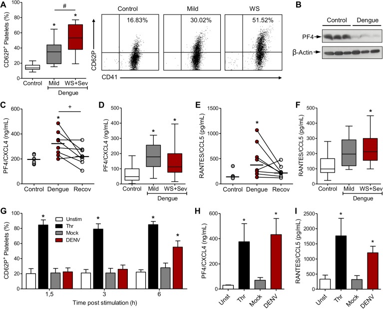
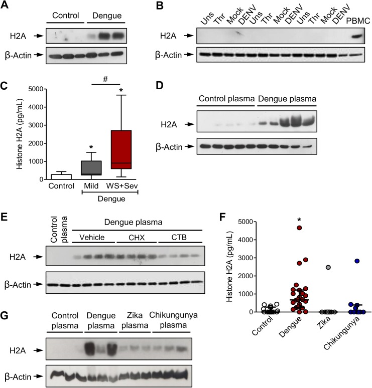
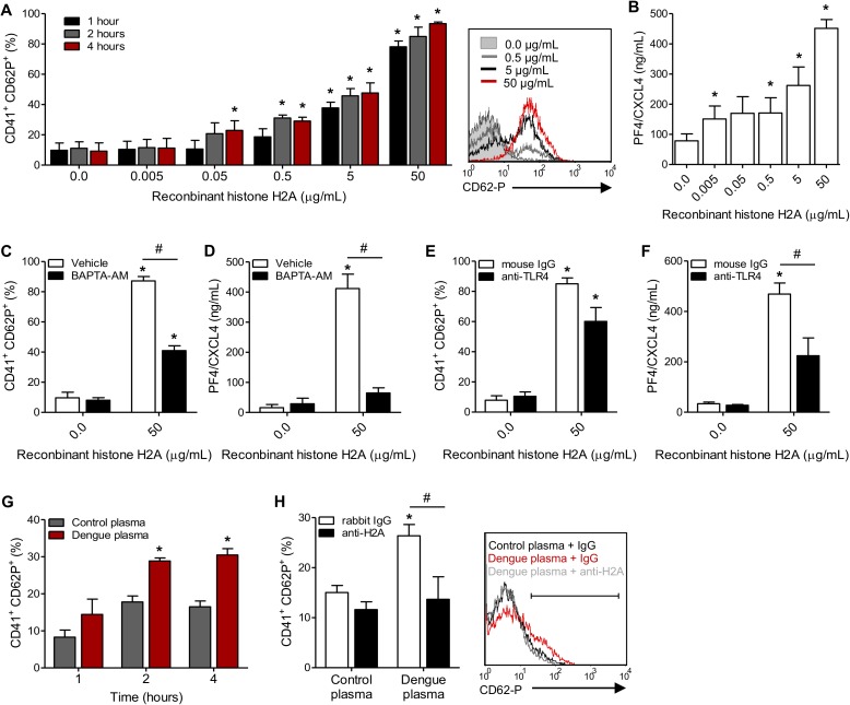
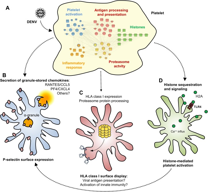
Dengue is the most prevalent human arbovirus disease worldwide. Dengue virus (DENV) infection causes syndromes varying from self-limiting febrile illness to severe dengue. Although dengue pathophysiology is not completely understood, it is widely accepted that increased inflammation plays important roles in dengue pathogenesis. Platelets are blood cells classically known as effectors of hemostasis which have been increasingly recognized to have major immune and inflammatory activities. Nevertheless, the phenotype and effector functions of platelets in dengue pathogenesis are not completely understood. Here we used quantitative proteomics to investigate the protein content of platelets in clinical samples from patients with dengue compared to platelets from healthy donors. Our assays revealed a set of 252 differentially abundant proteins. In silico analyses associated these proteins with key molecular events including platelet activation and inflammatory responses, and with events not previously attributed to platelets during dengue infection including antigen processing and presentation, proteasome activity, and expression of histones. From these results, we conducted functional assays using samples from a larger cohort of patients and demonstrated evidence for platelet activation indicated by P-selectin (CD62P) translocation and secretion of granule-stored chemokines by platelets. In addition, we found evidence that DENV infection triggers HLA class I synthesis and surface expression by a mechanism depending on functional proteasome activity. Furthermore, we demonstrate that cell-free histone H2A released during dengue infection binds to platelets, increasing platelet activation. These findings are consistent with functional importance of HLA class I, proteasome subunits, and histones that we found exclusively in proteome analysis of platelets in samples from dengue patients. Our study provides the first in-depth characterization of the platelet proteome in dengue, and sheds light on new mechanisms of platelet activation and platelet-mediated immune and inflammatory responses.


Conflict of interest statement

The authors declare no competing financial interests.

Figures

Fig 1
Fig 1. Distribution of mapped proteins in platelets from dengue patients and healthy volunteers.
(A) Venn diagram of mapped protein entries present in platelets from healthy volunteers (Control) and patients with dengue. Both conditions shared 2,557 proteins; 339 and 440 proteins were identified only in control or dengue platelet samples, respectively. (B) Volcano plot of all shared protein entries and their abundance in dengue and control conditions. The graph represents TFold pairwise analysis of the two biological conditions. Each dot represents a protein mapped according to its log2 (fold change) on the ordinate axis and its -log2 (t-test p-value) on the abscissa axis. Red dots indicate proteins that satisfy neither the fold-change cutoff nor the FDR cutoff α (0.05). Green dots depict protein entries that satisfy the fold-change cutoff but not FDR α. Orange dots indicate proteins that satisfy both fold-change and FDR α, but present low fold changes. Blue dots represent protein entries that satisfy all statistical filters. The result shows 86 proteins significantly up-regulated (blue dots above the dotted line on Volcano plot) and 81 proteins down-regulated (blue dots below the dotted line on the graph) in platelets from patients with dengue.
Fig 2
Fig 2. Interaction network of proteins differentially expressed between dengue and control platelets.
(A) List of all 905 interactions between 252 differentially abundant proteins detected by the proteomic analysis. (B) A GO analysis validated the following biological processes: “platelet activation”, “antigen processing and presentation”, “proteasome activity”, “inflammatory response” and “histones”. Proteins labeled with two colors (fill and boundary colors) are involved in two biological processes. Larger circles represent proteins upregulated in dengue. Small circles indicate proteins downregulated in dengue. Squares represent proteins detected only dengue (large) or control (small) conditions.
Fig 3
Fig 3. Dengue triggers platelet activation and chemokine secretion.
(A) P-selectin (CD62P) surface expression by platelets isolated from healthy volunteers (Control) and patients with mild dengue, dengue with warning signs (WS) and severe dengue (Sev). (percentage of positive cells by flow cytometry). Boxes indicate the median and interquartile ranges and whiskers indicate minimal and maximal values in each group. Representative dot plots are shown. (B) Western blot analysis of platelet factor 4 (PF4/CXCL4) and β-actin in platelets isolated from three control subjects and three patients with dengue. (C and E) PF4/CXCL4 and RANTES/CCL5 concentration in supernatants of platelets (1 x 10⁹ per mL) from 8 control subjects and 8 dengue patients that were cultured for 4h ex vivo. Platelets obtained from the same patients in the recovery phase (Recov) were also evaluated. Each dot represents the level of chemokines secreted by platelets from one dengue patient or control. Horizontal lines represent median. (D and F) Concentration of PF4/CXCL4 and RANTES/CCL5 in plasma from control subjects and patients with dengue. Boxes indicate the median and interquartile ranges and whiskers indicate minimal and maximal values in each group. (G-I) Platelets isolated from healthy volunteers were kept unstimulated (Unst) or stimulated with thrombin (Thr), DENV or Mock for the indicated times. Panel G shows the percentage of CD62P-expressing platelets, and panels H to I show the concentration of PF4/CXCL4 and RANTES/CCL5 in the supernatant of platelets incubated in each condition for 6h. Bars represent mean ± standard error of the mean of 4 to 8 independent experiments performed with platelets from individual donors.* means p<0.05 compared to Control, Unst or Mock; # indicates p<0.05 between patients with mild and WS+Sev dengue syndromes; + represents p<0.05 between dengue patients in the acute and recovery phase (paired t test).
Fig 4
Fig 4. Increased HLA class I on DENV-infected platelets depends on protein translation and proteasome activity.
(A) Western blot analysis for HLA class I and β-actin in freshly isolated platelets from two healthy control volunteers and two dengue-infected patients. (B-H) Platelets isolated from healthy volunteers were kept unstimulated (Unst) or stimulated with thrombin (Thr), DENV or Mock for the indicated times. Panel B shows the overall HLA class I expression in platelets from two independent donors at 6 hours post stimulation; and panel C shows the percent of platelets with high surface expression of HLA class I (HLA class IHigh) in each condition. (D-H) Platelets were exposed to DENV or Mock in the presence of DMSO (vehicle), bortezomib (1 μM) or cyclohexamide (10 μM). Panel D show the HLA class I expression at 6 hours post infection, panels E-F show the percent of HLA class I High expression and panels G-H depicts the P-selectin (CD62P) surface expression in platelets incubated in each condition. Bars represent mean ± standard error of the mean of 3 to 7 independent experiments from individual platelet donors. * indicates p<0.05 compared to unstimulated platelets or Mock; # means p<0.05 between platelets treated with Vehicle and Bortezomib or Cyclohexamide. Representative histograms are shown.
Fig 5
Fig 5. Platelets sequester circulating histone H2A in plasma from dengue-infected patients.
(A-B) Western blot analysis for histone H2A and β-actin in (A) freshly isolated platelets from three healthy volunteers (Control) and three patients with dengue; and in (B) platelets from three healthy volunteers that were kept unstimulated (Unst) or stimulated with thrombin (Thr), DENV or Mock for 6h. Human peripheral blood mononuclear cells (PBMC) were used as positive control for histone H2A expression. (C) Histone H2A concentration in plasma from control subjects or patients with mild dengue or dengue with warning signs and severe dengue (WS+Sev). Boxes indicate the median and interquartile ranges and whiskers indicate minimal and maximal values in each group. (D-E) Platelets were isolated from a healthy volunteer and incubated with 20% plasma from five dengue-infected patients (dengue plasma) or five healthy volunteers (control plasma) for 4 hours in the presence or absence of cyclohexamide (CHX), cytochalasin B (CTB) or DMSO (vehicle). (F) Histone H2A concentration in plasma from control subjects or patients with dengue, zika or chikungunya fever. Each dot represents the level of histone H2A in plasma from one patient or control. Lines represent median and interquartile range. (G) Western blot analysis for histone H2A and β-actin in platelets incubated with 20% plasma from three control subjects or three patients with dengue, zika or chikungunya. * means p<0.05 compared to control, zika or chikungunya; # indicates p<0.05 between patients with mild and WS+Sev dengue syndromes. Western blots (D, E and G) are representative of three independent experiments from individual platelet donors.
Fig 6
Fig 6. Circulating histone H2A in plasma from dengue-infected patients activates platelets.
(A-B) Platelets isolated from healthy volunteers were stimulated with recombinant human histone H2A at the indicated concentrations. (A) Platelet surface P-selectin (CD62-P) was evaluated 1, 2 and 4 hours post stimulation by flow cytometry and (B) PF4/CXCL4 concentration was measured in supernatants 4 hours post stimulation. (C-F) Surface P-selectin and PF4/CXCL4 concentration in the supernatants of platelets stimulated with recombinant histone H2A for 2 hour in the presence of (C-D) the calcium chelator BAPTA-AM (20 μM) or vehicle (DMSO); or (E-F) blocking antibody against TLR4 (20 μg/mL) or isotype matched IgG. (G-H) P-selectin expression on platelets exposed to (G) plasma from six dengue-infected patients (dengue plasma) or four heterologous healthy volunteers (control plasma) for the indicated time-points; and (H) platelets exposed to dengue plasma or control plasma for 4 hours in the presence of anti-histone H2A (20 μg/mL) or isotype matched IgG. Bars represent mean ± standard error of the mean of 3 independent experiments (A-F) and of 4 to 6 independent plasma donors (G-H). * indicates p<0.05 compared to control plasma or unstimulated platelets; # represents p<0.05 between platelets treated with BAPTA-AM and vehicle or plasma samples treated with anti-histone H2A or isotype matched IgG. Representative histograms (A and H) illustrate surface P-selectin on platelets four hours after stimulation.
Fig 7
Fig 7. Schematic representation of proteome changes in platelets from dengue-infected patients.
(A) Platelets from dengue-infected patients exhibit changes in their proteome, phenotype and function. Alterations in the platelet proteome in dengue appear to influence five main biological processes based on our analysis: (i) platelet activation; (ii) antigen processing and presentation; (iii) processes influenced by proteasome dependent protein catabolism; (iv) inflammatory response and; (v) histone expression and signaling. (B) DENV infection activates platelets triggering granule release of stored chemokines (i.e. PF4/CXCL4 and RANTES/CCL5) and P-selectin surface expression. (C) DENV also increases HLA class I protein expression and its surface presentation through mechanisms requiring proteasome protein processing. The nature of peptides processed by proteasome activity and presented in HLA class I remains unknown. (D) Platelets sequester circulating cell-free histone H2A from plasma, which contributes to platelet activation during dengue infection.

Similar articles

Cited by

References

    1. Bhatt S, Gething PW, Brady OJ, Messina JP, Farlow AW, et al. (2013) The global distribution and burden of dengue. Nature 496: 504–507. doi: 10.1038/nature12060 - DOI - PMC - PubMed
    1. Schmidt AC (2010) Response to dengue fever—the good, the bad, and the ugly? N Engl J Med 363: 484–487. doi: 10.1056/NEJMcibr1005904 - DOI - PubMed
    1. WHO (2009) Dengue: guidelines for diagnosis, treatment, prevention and control. - PubMed
    1. Vaughn DW, Green S, Kalayanarooj S, Innis BL, Nimmannitya S, et al. (1997) Dengue in the early febrile phase: viremia and antibody responses. J Infect Dis 176: 322–330. - PubMed
    1. Bozza FA, Cruz OG, Zagne SM, Azeredo EL, Nogueira RM, et al. (2008) Multiplex cytokine profile from dengue patients: MIP-1beta and IFN-gamma as predictive factors for severity. BMC Infect Dis 8: 86 doi: 10.1186/1471-2334-8-86 - DOI - PMC - PubMed