Review
. 2018 Mar;233(3):1897-1912.
doi: 10.1002/jcp.26017. Epub 2017 Jun 15.

Intimate connections: Inositol pyrophosphates at the interface of metabolic regulation and cell signaling

Affiliations
Review

Intimate connections: Inositol pyrophosphates at the interface of metabolic regulation and cell signaling

Stephen B Shears. J Cell Physiol. 2018 Mar.

Abstract

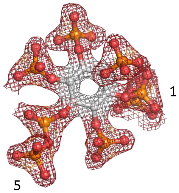
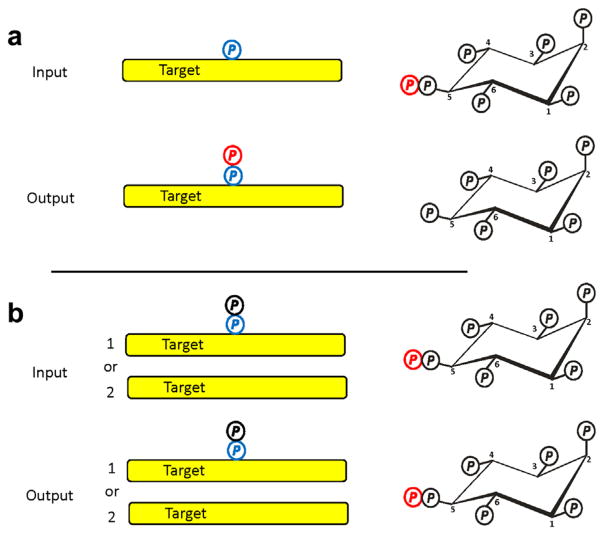
Inositol pyrophosphates are small, diffusible signaling molecules that possess the most concentrated three-dimensional array of phosphate groups in Nature; up to eight phosphates are crammed around a six-carbon inositol ring. This review discusses the physico-chemical properties of these unique molecules, and their mechanisms of action. Also provided is information on the enzymes that regulate the levels and hence the signaling properties of these molecules. This review pursues the idea that many of the biological effects of inositol pyrophosphates can be rationalized by their actions at the interface of cell signaling and metabolism that is essential to cellular and organismal homeostasis.

Keywords: cell signaling; inositol pyrophosphates; kinase; metabolism; phosphatase.


Figures

FIGURE 1
FIGURE 1
A cyclical pathway for PP-InsP turnover. Current evidence (described in the text) indicates that the pathway for PP-InsP turnover may be described as a cyclical interconversion of InsP6, 5-InsP7, 1,5-InsP8, and 1-InsP7, upon which is superimposed “futile” cycling between 5-InsP7 and 1,5-InsP8 (see text for details). IP6K, inositol hexakisphosphate kinase; PPIP5K, diphosphoinositolpentakisphosphate kinase; DIPP, diphosphoinositol polyphosphate phosphatase
FIGURE 2
FIGURE 2
Domain graphics of human PPIP5K1 and PPIP5K2. Domain graphics are shown for the human PPIP5K1 (Accession number BC057395.1) and PPIP5K2 (accession number XM_005271938); IDR, intrinsically disordered domain. The derivation of the amino-acid boundaries of each domain is as previously described (Gu et al., 2017). The % sequence identities across each of the three domains are provided
FIGURE 3
FIGURE 3
PPIP5Ks utilize a substrate capture site and a catalytic site for an unusual “catch-and-pass” reaction mechanism. (a) Electrostatic surface rendering of the structure of the kinase domain of human PPIP5K2. Blue represents electropositive, and red indicates electronegative. A magnified image of the catalytic site is also shown. Note that two near parallel grooves form a “staggered-H” that ensures only InsP6 or 5-InsP7 may enter the active site and be phosphorylated. (b) Surface representation of PPIP5K highlighting the binding of a 5-phosphonoacetate analogue of 5-InsP7 to both a surface mounted “substrate capture” site and a catalytic site (note that steric constraints prevent both sites from being occupied simultaneously) (Wang, Godage, et al., 2014). (c) Depiction of the molecular gymnastics required for delivery of substrate between the two sites: a 100° ring flip and a lateral movement (broken yellow line) of 7 Å (Wang, Godage, et al., 2014). (d) To avoid the sterically restricted active site constraining catalytic activity, does substrate in the bulk phase (the ping-pong ball) have an increased probability of being appropriately oriented to enter the active site (the neck of the goldfish bowl) because of the proximal substrate capture site (the green cone)?
FIGURE 4
FIGURE 4
Phosphorylation of Ins(1,3,4,5,6)P5 by IP6Ks. The figure summarizes data obtained in vitro which demonstrate that two PP-InsPs can be generated by IP6K-mediated phosphorylation of Ins(1,3,4,5,6)P5 (Draskovic et al., 2008)
FIGURE 5
FIGURE 5
Electron density map of 1,5-InsP8. 1,5-InsP8 is depicted as a refined 2 Fo-Fc map contoured at 2.0 ςthat is shown in blue mesh. This information was captured from crystals of the kinase domain of PPIP5K2 that also contained ADP (Capolicchio et al., 2014). The 1- and 5-diphosphate groups are numbered
FIGURE 6
FIGURE 6
Assessing the status of protein pyrophosphorylation in vivo by a back-pyrophosphorylation assay in vitro. (a) Idealized assay input and output are shown for an in vitro, back-phosphorylation assay. The input is 5-β-32P-InsP7 and a purified target protein that, in vivo, had been phosphorylated by CK2 (blue “P”), but not pyrophosphorylated by a PP-InsP. The assay “output” is pyrophosphorylated target (red “P”) and InsP6. The conclusion to be drawn from this outcome is that the target was not pyrophosphorylated in vivo. (b) Describes two different assay inputs that both lead to an assay output showing no protein pyrophosphorylation: condition 1 is target pyrophosphorylation in vivo; condition 2 is no target phosphorylation by CK2 in vivo. Thus, the assay output data are ambiguous as to the phosphorylation status of the target in vivo. This problem can be resolved with a second, independent back-phosphorylation assay in vitro, using CK2. The latter will only phosphorylate the target if condition 2 is satisfied. The absence of CK2-mediated phosphorylation in vitro would support condition 1: target pyrophosphorylation in vivo

References

    1. Aird KM, Worth AJ, Snyder NW, Lee JV, Sivanand S, Liu Q, … Zhang R. ATM couples replication stress and metabolic reprogramming during cellular senescence. Cell Reports. 2015;11:893–901. - PMC - PubMed
    1. Azevedo C, Burton A, Ruiz-Mateos E, Marsh M, Saiardi A. Inositol pyrophosphate mediated pyrophosphorylation of AP3B1 regulates HIV-1 Gag release. Proceedings of the National Academy of Sciences of the United States of America. 2009;106:21161–21166. - PMC - PubMed
    1. Azevedo C, Saiardi A. Eukaryotic phosphate homeostasis: The inositol pyrophosphate perspective. Trends in Biochemical Sciences. 2017;42:219–231. - PubMed
    1. Banfic H, Bedalov A, York JD, Visnjic D. Inositol pyrophosphates modulate S phase progression after pheromone-induced arrest in Saccharomyces cerevisiae. Journal of Biological Chemistry. 2013;288:1717–1725. - PMC - PubMed
    1. Bao Y, Ledderose C, Seier T, Graf AF, Brix B, Chong E, Junger WG. Mitochondria regulate neutrophil activation by generating ATP for autocrine purinergic signaling. Journal of Biological Chemistry. 2014;289:26794–26803. - PMC - PubMed