. 2017 Nov;37(11):1325-1332.
doi: 10.1002/jat.3462. Epub 2017 May 24.

Mechanism of graphene-induced cytotoxicity: Role of endonucleases

Affiliations

Mechanism of graphene-induced cytotoxicity: Role of endonucleases

Tariq Fahmi et al. J Appl Toxicol. 2017 Nov.

Abstract

Graphene, a crystalline allotrope or carbon, presents numerous useful properties; however, its toxicity is yet to be determined. One of the most dramatic and irreversible toxic abilities of carbon nanomaterials is the induction of DNA fragmentation produced by endogenous cellular endonucleases. This study demonstrated that pristine graphene exposed to cultured kidney tubular epithelial cells is capable of inducing DNA fragmentation measured by terminal deoxynucleotidyl transferase dUTP nick end labeling (TUNEL) assay, which is usually associated with cell death. TUNEL (cell death) and endonuclease activity measured using a near infrared fluorescence probe was significantly higher in cells containing graphene aggregates detected by Raman spectroscopy. The elevation of TUNEL coincided with the increased abundance of heme oxygenase 1 (HO-1), heat shock protein 90 (HSP90), active caspase-3 and endonucleases (deoxyribonuclease I [DNase I] and endonuclease G [EndoG]), as measured by quantitative immunocytochemistry. Specific inhibitors for HO-1, HSP90, caspase-3, DNase I and EndoG almost completely blocked the DNA fragmentation induced by graphene exposure. Therefore, graphene induces cell death through oxidative injury, caspase-mediated and caspase-independent pathways; and endonucleases DNase I and EndoG are important for graphene toxicity. Inhibition of these pathways may ameliorate cell injury produced by graphene. Copyright © 2017 John Wiley & Sons, Ltd.

Keywords: DNA fragmentation; DNase I; EndoG; cell death; endonucleases; graphene; oxidative injury.


Conflict of interest statement

Conflict of interest

The authors did not report any conflict of interest.

Figures

Figure 1.
Figure 1.
Measurement of graphene toxicity in renal tubular epithelial cells. (A) Nuclear TUNEL staining (green) indicative of DNA fragmentation and cell death is located in the cell that contains a graphene cluster (shown by red arrow). (B) TUNEL assay measurement of graphene cytotoxicity at varying concentrations. (C) Quantification of DNase activity near infrared fluorescence. (D) Nuclear polymeric DNA staining by DAPI in NRK-52E cells. C, control vehicle-treated cells; Gn, cells exposed to graphene; UT, untreated cells.
Figure 2.
Figure 2.
Cells containing graphene clusters have more DNA fragmentation. (A) Representative 2D Raman maps developed based on the graphene specific signature (left). Integrated Raman spectroscopy signal is higher in TUNEL+ versus TUNEL cells (right). (B) DNA endonuclease activity measured by NIRF probe signal directly correlates with graphene quantity (mean intensity). (C) DNA quantity measure by DAPI mean intensity reversely correlates with graphene quantity (mean intensity), which indicates degradation and removal of some DNA. NIRF, near infrared fluorescence.
Figure 3.
Figure 3.
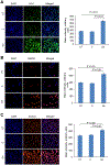
Immunocytochemical measurement of oxidative and apoptotic markers affected by graphene (50 μg ml−1) exposure of NRK-52E cells. (A) Graphene induces HO-1 (green staining). (B–D) HSP90, EndoG and active caspase-3 immunostaining (red color) and quantification of their mean intensities, respectively, in NRK-52E cells. (E,F) Quantification of DNase I and CAD mean intensities, respectively, in NRK-52E cells exposed to graphene. C, control vehicle-treated cells; Gn, cells exposed to graphene; HO-1, heme-oxygenase-1; HSP90, heatshock protein 90; UT, untreated cells.
Figure 3.
Figure 3.
Immunocytochemical measurement of oxidative and apoptotic markers affected by graphene (50 μg ml−1) exposure of NRK-52E cells. (A) Graphene induces HO-1 (green staining). (B–D) HSP90, EndoG and active caspase-3 immunostaining (red color) and quantification of their mean intensities, respectively, in NRK-52E cells. (E,F) Quantification of DNase I and CAD mean intensities, respectively, in NRK-52E cells exposed to graphene. C, control vehicle-treated cells; Gn, cells exposed to graphene; HO-1, heme-oxygenase-1; HSP90, heatshock protein 90; UT, untreated cells.
Figure 4.
Figure 4.
Alleviation of graphene toxicity by using chemical inhibitors after 48 h exposure of NRK-52E cells with graphene (50 μg ml−1). Quantification of TUNEL-positive cells in NRK-52E cells treated with graphene in the presence of chemical inhibitors for HO-1, HSP90, caspase 3, DNase I and EndoG (p < 0.01 for all bars vs. Gn).

References

    1. Ahamed M, Alsalhi MS, Siddiqui MK. 2010. Silver nanoparticle applications and human health. Clin. Chim. Acta 411: 1841–1848. - PubMed
    1. Akhavan O, Ghaderi E. 2010. Toxicity of graphene and graphene oxide nanowalls against bacteria. ACS Nano 4: 5731–5736. - PubMed
    1. Apostolov EO, Soultanova I, Savenka A, Bagandov OO, Yin X, Stewart AG, Walker RB, Basnakian AG. 2009. Deoxyribonuclease I is essential for DNA fragmentation induced by gamma radiation in mice. Radiat. Res 172: 481–492. - PMC - PubMed
    1. Basnakian AG, Kaushal GP, Shah SV. 2002. Apoptotic pathways of oxidative damage to renal tubular epithelial cells. Antioxid. Redox Signal 4: 915–924. - PubMed
    1. Buzder T, Yin X, Wang X, Banfalvi G, Basnakian AG. 2009. Uptake of foreign nucleic acids in kidney tubular epithelial cells deficient in proapoptotic endonucleases. DNA Cell Biol. 28: 435–442. - PMC - PubMed

MeSH terms

LinkOut - more resources