. 2017 Aug;65(8):1264-1277.
doi: 10.1002/glia.23160. Epub 2017 May 22.

Degradation of heme oxygenase-1 by the immunoproteasome in astrocytes: A potential interferon-γ-dependent mechanism contributing to HIV neuropathogenesis

Affiliations

Degradation of heme oxygenase-1 by the immunoproteasome in astrocytes: A potential interferon-γ-dependent mechanism contributing to HIV neuropathogenesis

Colleen E Kovacsics et al. Glia. 2017 Aug.

Abstract

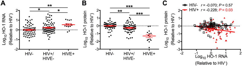
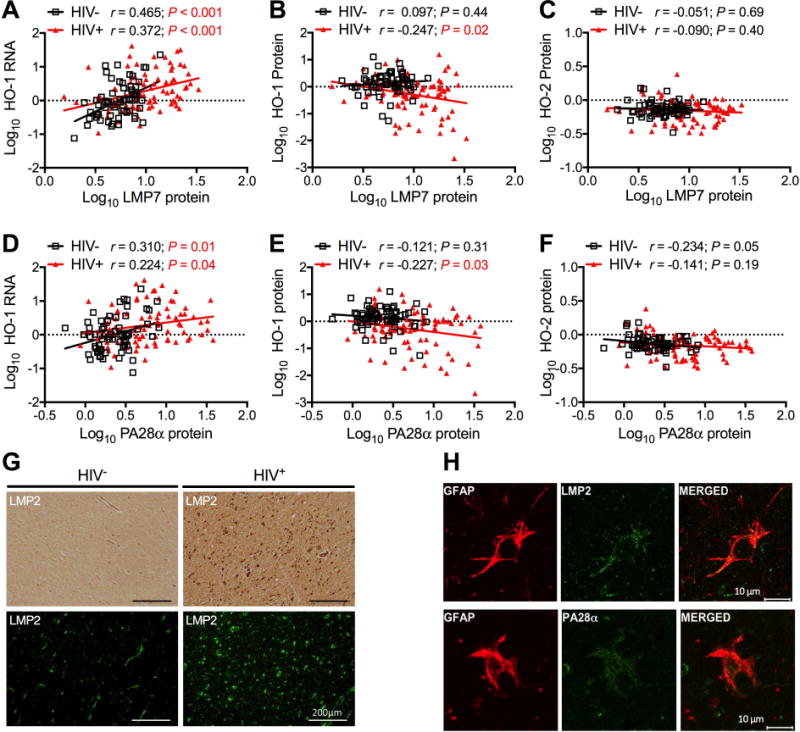
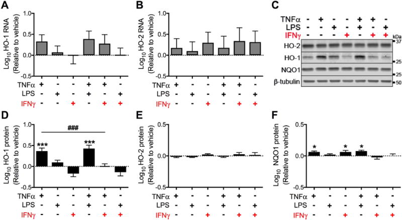
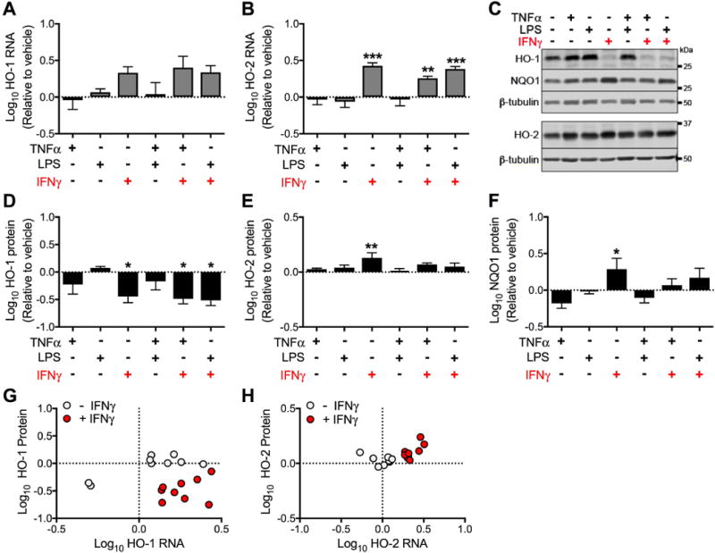
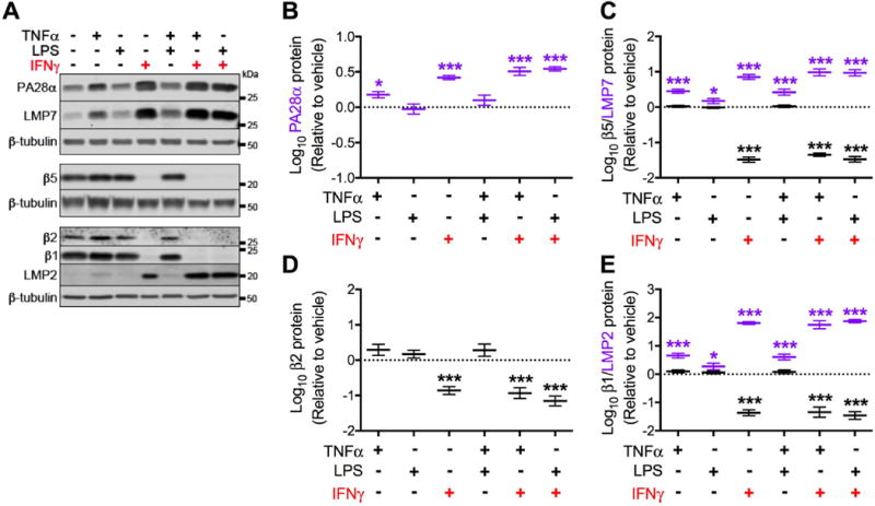
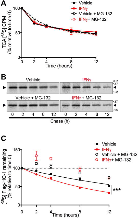
Induction of the detoxifying enzyme heme oxygenase-1 (HO-1) is a critical protective host response to cellular injury associated with inflammation and oxidative stress. We previously found that HO-1 protein expression is reduced in brains of HIV-infected individuals with HIV-associated neurocognitive disorders (HAND) and in HIV-infected macrophages, where this reduction associates with enhanced glutamate release and neurotoxicity. Because HIV-infected macrophages are a small component of the cellular content of the brain, the reduction of macrophage HO-1 expression likely accounts for a small portion of brain HO-1 loss in HIV infection. We therefore investigated the contribution of astrocytes, the major pool of brain HO-1. We identified immunoproteasome-mediated HO-1 degradation in astrocytes as a second possible mechanism of brain HO-1 loss in HIV infection. We demonstrate that prolonged exposure of human fetal astrocytes to interferon-gamma (IFNγ), an HIV-associated CNS immune activator, selectively reduces expression of HO-1 protein without a concomitant reduction in HO-1 RNA, increases expression of immunoproteasome subunits, and decreases expression of constitutive proteasome subunits, consistent with a shift towards increased immunoproteasome activity. In HIV-infected brain HO-1 protein reduction also associates with increased HO-1 RNA expression and increased immunoproteasome expression. Finally, we show that IFNγ treatment of astrocytic cells reduces HO-1 protein half-life in a proteasome-dependent manner. Our data thus suggest unique causal links among HIV infection, IFNγ-mediated immunoproteasome induction, and enhanced HO-1 degradation, which likely contribute to neurocognitive impairment in HAND. Such IFNγ-mediated HO-1 degradation should be further investigated for a role in neurodegeneration in inflammatory brain conditions.

Brief summary: Kovacsics et al. identify immunoproteasome degradation of heme oxygenase-1 (HO-1) in interferon gamma-stimulated astrocytes as a plausible mechanism for the observed loss of HO-1 protein expression in the brains of HIV-infected individuals, which likely contributes to the neurocognitive impairment in HIV-associated neurocognitive disorders.

Keywords: HAND; HIV-associated neurocognitive disorders; immunoproteasome; interferon gamma.


Conflict of interest statement

Conflict of Interest Statement

The other authors have no conflicts of interest.

Figures

Figure 1
Figure 1. HO-1 RNA is elevated and HO-1 protein is reduced in the dorsolateral prefrontal cortex of HIV-infected individuals
(A) HO-1 RNA expression in the DLPFC determined by real-time quantitative PCR. HIV− = (HIV-negative, n=66); HIV+/HIVE− = (HIV-positive without HIV encephalitis, n = 76); and HIVE = (HIV-positive with HIV encephalitis, n = 14). (B) HO-1 protein expression determined by Western blot in 66 HIV−, 75 HIV+/HIVE−, and 14 HIVE brain tissue samples. Fold-change in expression was calculated relative to the average of HIV− groups after normalization to GAPDH (RNA) or β-tubulin (protein). Data were log transformed and mean HIV− group value set to 0 (dotted line). Solid black lines indicate mean ± SEM. Groups were analyzed by ANOVA with post hoc Holm-Sidak test. *P < 0.05; **P < 0.01; ***P < 0.001. (C) Correlation between HO-1 RNA and protein expression in 65 HIV− and 89 HIV+ (75 HIV+/HIV− and 14 HIVE) was determined by Pearson’s correlation with line of best fit determined by linear regression.
Figure 2
Figure 2. HO-1 protein loss is associated with immunoproteasome induction in HIV-infected brain
Expression of immunoproteasome subunits LMP7 (A, B, C) and PA28α (D, E, F) was determined by Western blot in DLPFC samples. Immunoproteasome expression was correlated with (A, D) HO-1 RNA in 64 HIV and 87 HIV+ samples and with (B, E) HO-1 and (C, F) HO-2 protein in 65 HIV and 88 HIV+ DLPFC samples. Correlations were determined by Pearson’s correlation with line of best fit determined by linear regression. (G) Immunoproteasome subunit LMP2 was visualized in subcortical white matter by immunohistochemistry (top panels) and immunofluorescence (bottom panels). Scale bar = 200 μm. (H) Immunoproteasome subunits were localized to astrocytes by dual indirect immunofluorescence staining of LMP2 (top panels) or PA28α (bottom panels) with the astrocyte marker GFAP in subcortical white matter of HIVE cases. Scale bar =10 μm.
Figure 3
Figure 3. Acute (24 hours) exposure to IFNγ does not significantly reduce HO-1 expression in astrocytes
Primary human fetal astrocytes were exposed to TNFα, LPS, and IFNγ (alone or in combination) for 24 hours. (A) HO-1 and (B) HO-2 RNA expression was determined by real-time quantitative PCR. Fold change in expression was calculated relative to vehicle after normalization to ACTB (β-Actin). (C) Representative Western blot from a single biological replicate. Quantification of (D) HO-1, (E) HO-2 and (F) NQO1 protein expression relative to vehicle after normalization to β-tubulin. Data were log transformed and values represent mean ± SEM (n = 4 biological replicates) of the fold change in expression from vehicle (dotted line). Statistical comparisons were made by RM-ANOVA with post hoc Holm-Sidak test. *P < 0.05; ***P < 0.001; ****P < 0.0001 vs. vehicle. ###P < 0.001 for indicated comparison.
Figure 4
Figure 4. Prolonged (15 days) exposure to IFNγ significantly reduces HO-1 protein expression, but not RNA expression in astrocytes
Primary human fetal astrocytes were exposed to TNFα, LPS, and IFNγ (alone or in combination) for 15 days. Media and treatments were replaced every 3 days. (A) HO-1 and (B) HO-2 RNA expression was determined by real-time quantitative PCR. Fold change in expression was calculated relative to vehicle after normalization. (C) Representative Western blot from a single biological replicate. Quantification of (D) HO-1, (E) HO-2 and (F) NQO1 protein expression relative to vehicle after normalization. Data were log transformed and values represent mean ± SEM (n = 4 biological replicates) of the fold change in expression from vehicle (dotted line). Statistical comparisons to vehicle were made by RM-ANOVA with post hoc Holm-Sidak test. *P < 0.05; **P < 0.01; ***P < 0.001. Fold change in RNA and protein expression of (G) HO-1 and (H) HO-2 were individually plotted for each treatment condition from (n = 3 biological replicates, treatments and protein and RNA extraction occured in parallel wells). Solid black circles represent TNFα, LPS, and TNFα + LPS; solid red circles represent IFNγ, IFNγ + TNFα, and IFNγ + LPS.
Figure 5
Figure 5. Prolonged (15 days) exposure to IFNγ significantly induces immunoproteasome subunit expression and decreases constitutive proteasome subunit expression in astrocytes
Primary human fetal astrocytes were exposed to TNFα, LPS, and IFNγ (alone or in combination) for 15 days. Media and treatments were replaced every 3 days. (A) Representative Western blot of immunoproteasome (PA28α, LMP7, LMP2) and constitutive proteasome (β5, β2, β1) subunits from a single biological replicate. Quantification of (B) PA28α, (C) LMP7 and β5, (D) β2 and (E) LMP2 and β1 protein expression relative to vehicle after normalization. Data were log transformed and values represent mean ± SEM (n = 4 biological replicates) of the fold change from vehicle (dotted line). Statistical comparisons to vehicle were made by RM-ANOVA with post hoc Holm-Sidak test. *P < 0.05; ***P < 0.001; **** P < 0.0001.
Figure 6
Figure 6. IFNγ effects on expression of proteasome subunits and HO-1 are dose-dependent
Primary human fetal astrocytes were exposed to IFNγ (0.1 pg/mL – 25 ng/mL) for 15 days. Media and treatments were replaced every 3 days. Western blot densitometry quantification of (A) HO-1, (B) PA28α, (C) LMP7, and β5 protein expression relative to vehicle after normalization to β-tubulin. Data were log transformed and values represent mean ± SEM (n = 4 biological replicates) of the fold change from vehicle (dotted line). Statistical comparisons to vehicle were made by RM-ANOVA with post hoc Holm-Sidak test. *P < 0.05; **P < 0.01; ***P < 0.001; **** P < 0.0001. Statistical comparisons between consecutive IFNγ doses were made by RM-ANOVA with post hoc Holm-Sidak test. #P < 0.05; ##P < 0.01; ###P < 0.001; ####P < 0.0001.
Figure 7
Figure 7. IFNγ increases the rate of proteasome-dependent HO-1 degradation
U251 cells transiently expressing FLAG-HO-1 and exposed to IFNγ (10 ng/mL) for 3 days were used in pulse chase assays to measure HO-1 degradation. Cells were pulse labeled with [35S]methionine/cysteine and chased for indicated time points. Degradation mediated by proteasome activity was assessed by treating cells 2 hours prior to labeling with the proteasome inhibitor MG-132 (5 μM) or DMSO (vehicle). (A) Intracellular protein degradation was determined by [35S] liquid scintillation counting of TCA-precipitable lysate fractions. Radioactivity was measured as counts per minute (CPM) and are expressed as a percentage relative to time 0 within each independent experiment (n=3). Data represent mean ± SEM. Statistical analysis of treatment conditions at each time point was performed by two-way RM-ANOVA with post-hoc Holm-Sidak test. (B) Representative phosphorimage used for densitometric quantification of radiolabeled FLAG-HO-1. Black arrows indicate radiolabeled FLAG-HO-1. (C) Degradation of HO-1 protein was determined by densitometric quantification of radiolabeled FLAG-HO-1 and are expressed as a percentage relative to time 0 within each independent experiment (n=3). Data represent mean ± SEM. Protein half-life in vehicle and IFNγ conditions (closed circles) was determined by one-phase exponential decay and rate of decay (k) was compared by Extra sum of squares F test. ***P < 0.001. The effects of MG-132 at each time point was analyzed by two-way RM-ANOVA with post hoc Holm-Sidak test (significance not shown in figure). IFNγ vs. IFNγ+MG-132: P < 0.001 at all time points. PBS vs. PBS + MG-132: P < 0.01 at 8hr; P < 0.001 at 2, 4, and 12 hr.

References

    1. Aki M, Shimbara N, Takashina M, Akiyama K, Kagawa S, Tamura T, Tanahashi N, Yoshimura T, Tanaka K, Ichihara A. Interferon-gamma induces different subunit organizations and functional diversity of proteasomes. J Biochem. 1994;115:257–69. - PubMed
    1. Akiyama K, Yokota K, Kagawa S, Shimbara N, Tamura T, Akioka H, Nothwang HG, Noda C, Tanaka K, Ichihara A. cDNA cloning and interferon gamma down-regulation of proteasomal subunits X and Y. Science. 1994;265:1231–4. - PubMed
    1. Alam J, Cook JL. How many transcription factors does it take to turn on the heme oxygenase-1 gene? Am J Respir Cell Mol Biol. 2007;36:166–74. - PubMed
    1. Amadio M, Scapagnini G, Davinelli S, Calabrese V, Govoni S, Pascale A. Involvement of ELAV RNA-binding proteins in the post-transcriptional regulation of HO-1. Front Cell Neurosci. 2014;8:459. - PMC - PubMed
    1. Ambegaokar SS, Kolson DL. Heme oxygenase-1 dysregulation in the brain: implications for HIV-associated neurocognitive disorders. Curr HIV Res. 2014;12:174–88. - PMC - PubMed

Publication types

MeSH terms