

Clinical and laboratory phenotype variability in type 2M von Willebrand disease
- PMID: 28544236
- PMCID: PMC5538962
- DOI: 10.1111/jth.13742
Clinical and laboratory phenotype variability in type 2M von Willebrand disease
Abstract
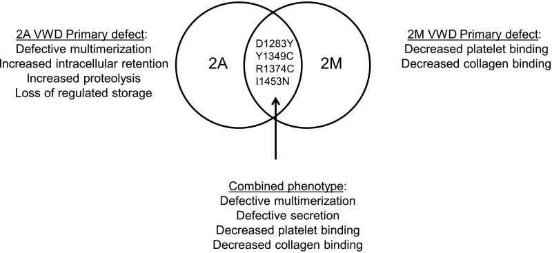
Essentials The pathophysiology of type 2M von Willebrand disease (VWD) is poorly understood. Sequence variations in type 2M VWD subjects were characterized. A high degree of clinical and laboratory variability exists within type 2M VWD variants. Some type 2M variants may share features of type 2A VWD.
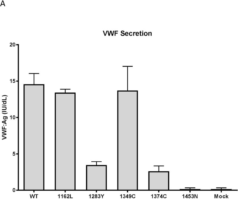
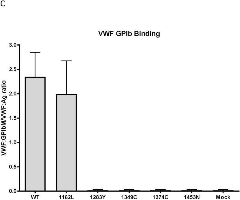
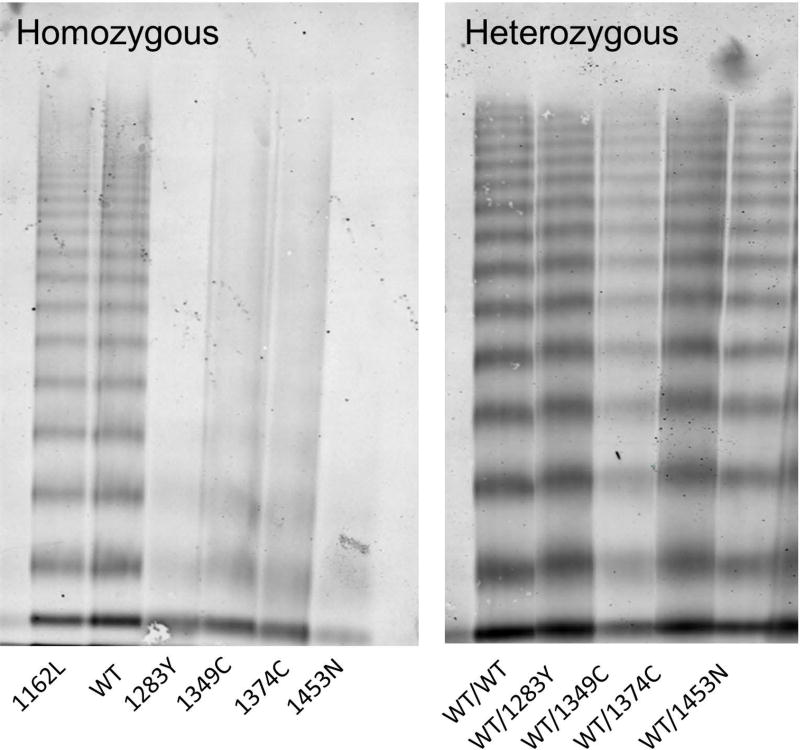
Summary: Background von Willebrand factor (VWF) is a multimeric coagulation factor that tethers platelets to injured subendothelium. Type 2M von Willebrand disease (VWD) is characterized by a qualitative defect in VWF with preserved multimer distribution. Objectives Through the Zimmerman Program for the Molecular and Clinical Biology for VWD, five VWF sequence variations were studied in subjects diagnosed with type 2M VWD. Methods Bleeding phenotype was assessed using the ISTH bleeding assessment tool. Full-length VWF gene sequencing was performed for each subject. Each variant was placed into a recombinant VWF vector using site-directed mutagenesis and expressed in HEK293T cells as homozygous or heterozygous VWF. Variant expression, collagen binding and platelet GPIbα binding were studied through ELISA assays. Multimer analysis was performed by gel electrophoresis. Results Bleeding scores were elevated for all subjects except for the p.P1162L and p.R1374C variants. Although all had reduced VWF ristocetin cofactor activity/VWF antigen ratios on plasma testing, recombinant VWF did not show a classic type 2M phenotype for any of the five variants. Homozygous expression of variants p.D1283Y, p.R1349C, p.R1374C and p.I1453N was consistent with type 2A VWD, although all had normal expression as heterozygous recombinant VWF. Variant p.P1162L had normal VWF expression and function, consistent with the lack of bleeding symptoms. Conclusions Although originally classified as type 2M VWD, these homozygous recombinant VWF variants do not fulfill complete 2M VWD diagnostic criteria. A better classification schema and improved testing for putative type 2M variants is needed in order to effectively diagnose and treat affected patients.
Keywords: clinical laboratory techniques; hemorrhage; platelets; von Willebrand disease; von Willebrand factor.
© 2017 International Society on Thrombosis and Haemostasis.
Conflict of interest statement
L. N. Boggio has served as a consultant for Alnylam, Bayer, Biogen, CSL Behring, Grifols, Novo Nordisk, Octapharma, OPKO, Pfizer, and Shire. S. R. Lentz. has received grants from NIH. V. H. Flood has received grants from NIH and has served as a consultant for CSL Behring and Shire.
Figures


References
-
- Fujimura Y, Titani K, Holland LZ, Russell SR, Roberts JR, Elder JH, Ruggeri ZM, Zimmerman TS. Von willebrand factor. A reduced and alkylated 52/48-kDa fragment beginning at amino acid residue 449 contains the domain interacting with platelet glycoprotein ib. J Biol Chem. 1986;261:381–5. - PubMed
-
- Pareti FI, Niiya K, McPherson JM, Ruggeri ZM. Isolation and characterization of two domains of human von willebrand factor that interact with fibrillar collagen types I and III. J Biol Chem. 1987;262:13835–41. - PubMed
-
- Sadler JE, Budde U, Eikenboom JC, Favaloro EJ, Hill FG, Holmberg L, Ingerslev J, Lee CA, Lillicrap D, Mannucci PM, Mazurier C, Meyer D, Nichols WL, Nishino M, Peake IR, Rodeghiero F, Schneppenheim R, Ruggeri ZM, Srivastava A, Montgomery RR, Federici AB. Working Party on von Willebrand Disease Classification. Update on the pathophysiology and classification of von willebrand disease: A report of the subcommittee on von willebrand factor. J Thromb Haemost. 2006;4:2103–14. - PubMed
-
- Nichols WL, Hultin MB, James AH, Manco-Johnson MJ, Montgomery RR, Ortel TL, Rick ME, Sadler JE, Weinstein M, Yawn BP. Von willebrand disease (VWD): Evidence-based diagnosis and management guidelines, the national heart, lung, and blood institute (NHLBI) expert panel report (USA) Haemophilia. 2008;14:171–232. - PubMed
Publication types
MeSH terms
Substances
Grants and funding
LinkOut - more resources
Full Text Sources
Other Literature Sources
Medical
Miscellaneous
