. 2017 Sep;105(9):2562-2571.
doi: 10.1002/jbm.a.36113. Epub 2017 Jun 15.

Galectin-1 promotes an M2 macrophage response to polydioxanone scaffolds

Affiliations

Galectin-1 promotes an M2 macrophage response to polydioxanone scaffolds

Daniel Abebayehu et al. J Biomed Mater Res A. 2017 Sep.

Abstract

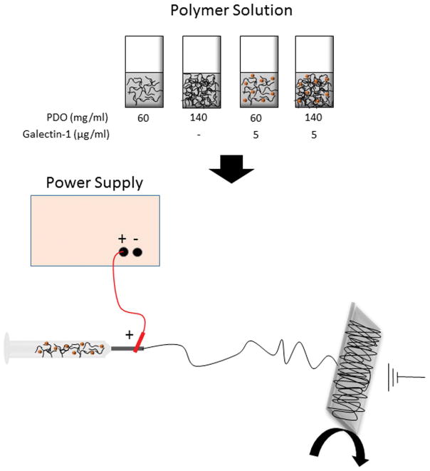
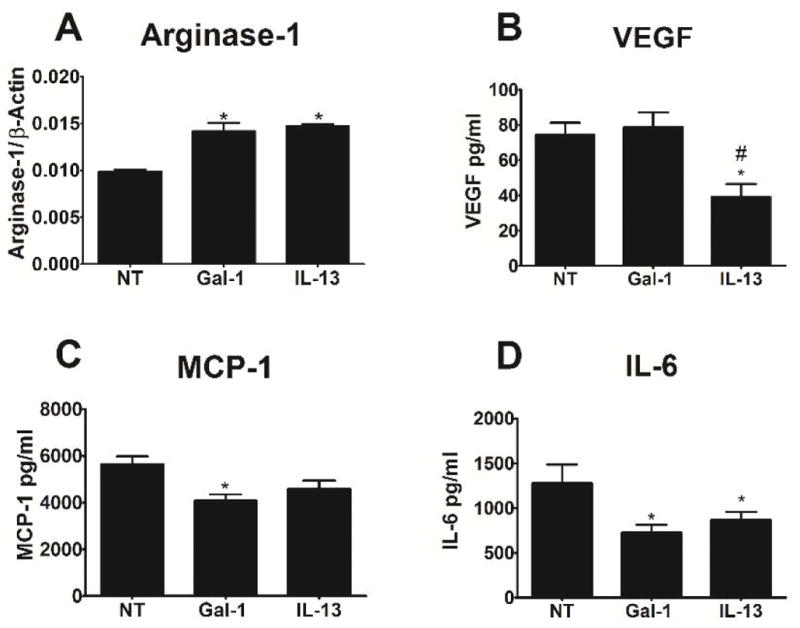
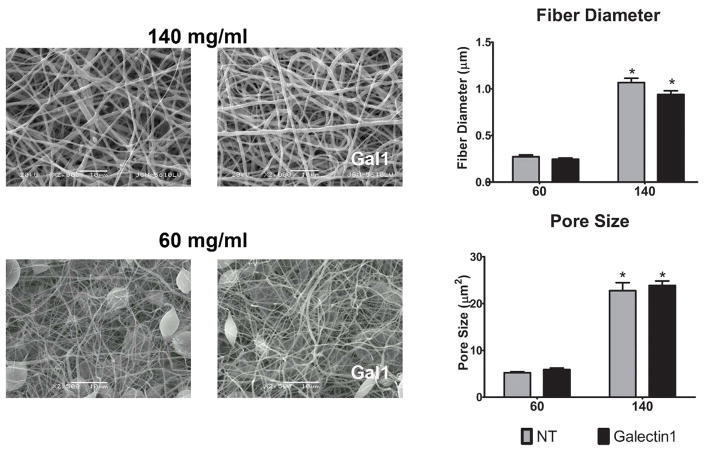
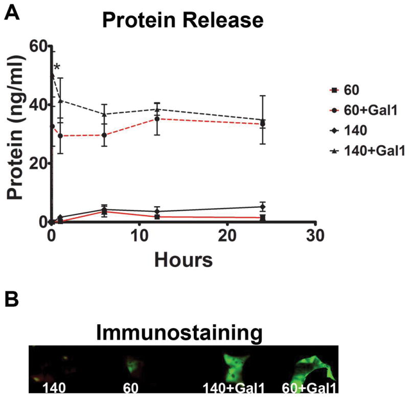
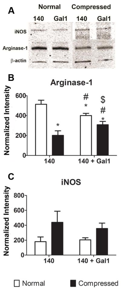
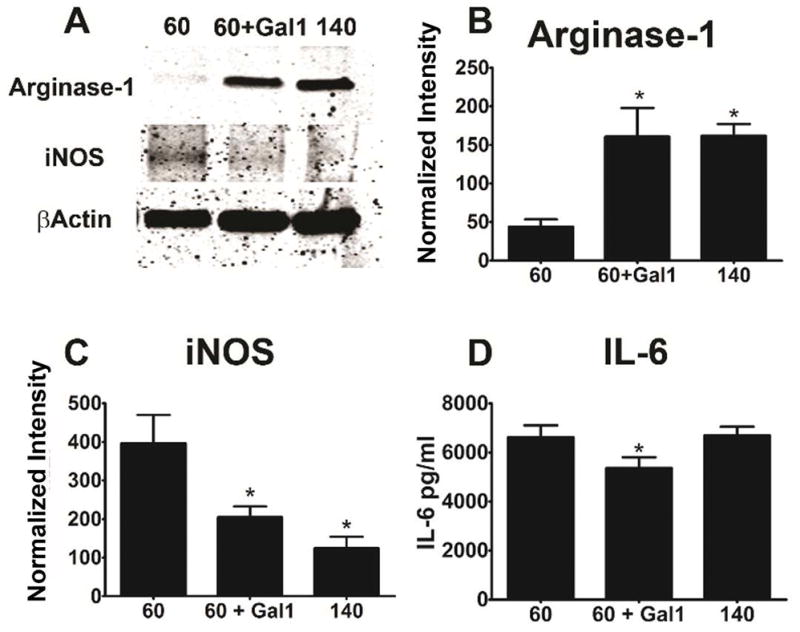
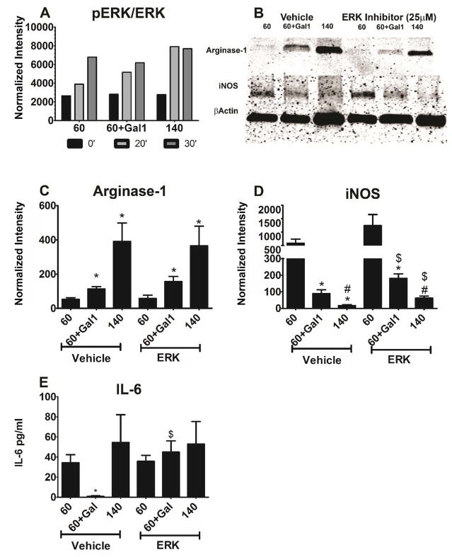
Regulating soft tissue repair to prevent fibrosis and promote regeneration is central to creating a microenvironment conducive to soft tissue development. Macrophages play an important role in this process. The macrophage response can be modulated using biomaterials, altering cytokine and growth factor secretion to promote regeneration. Electrospun polydioxanone (PDO) fiber scaffolds promoted an M2 phenotype when macrophages were cultured on large diameter, highly porous scaffolds, but an M1 phenotype on smaller diameter fibers. In this study, we investigated whether incorporation of galectin-1, an immunosuppressive protein that enhances muscle regeneration, could promote the M2 response. Galectin-1 was incorporated into large and small fiber PDO scaffolds during electrospinning. Galectin-1 incorporation increased arginase-1 and reduced iNOS and IL-6 production in mouse bone-marrow derived macrophages compared with PDO alone for both scaffold types. Inhibition of ERK mitogen-activated protein kinase did not alter galectin-1 effects on arginase-1 and iNOS expression, but reversed IL-6 suppression, indicating that IL-6 is mediated by a different mechanism. Our results suggest that galectin-1 can be used to modulate macrophage commitment to a pro-regenerative M2 phenotype, which may positively impact tissue regeneration when using small diameter PDO scaffolds. © 2017 Wiley Periodicals, Inc. J Biomed Mater Res Part A: 105A: 2562-2571, 2017.

Keywords: galectin-1; immunomodulation; macrophage; polydioxanone.


Figures

Figure 1
Figure 1. Schematic of method used to produce electrospun fibers with galectin-1
Figure 2
Figure 2. Galectin-1 promotes arginase and reduces IL-6 cytokine production similar to the effects of IL-13
Bone marrow-derived macrophages were cultured in media containing soluble galectin-1 (Gal1), IL-13, or no further treatment (NT) for 1 day. (A) Arginase-1 was measured by western blotting. (B–D) VEGF, MCP-1, and IL-6 were measured by ELISA. * indicates a difference from no treatment (p < 0.05). Data shown are means ± SEM of 6 samples from a representative experiment. Experiments were repeated to ensure validity.
Figure 3
Figure 3. Fiber diameter and pore size are unaffected by galectin-1 incorporation
Representative scanning electron micrographs of 140 mg/ml and 60 mg/ml electrospun scaffolds incorporating with 5 μg/ml galectin-1. Bar charts show the diameter and pore size measurements of 60 fibers from 3 samples. * indicates a difference when comparing 140 mg/ml scaffolds to the corresponding 60 mg/ml scaffold (p < 0.05).
Figure 4
Figure 4. Galectin-1 release
Protein levels were measured at 0, 0.25, 1, 6, 12, and 24 hours using 140 mg/ml and 60 mg/ml electrospun PDO scaffolds with galectin-1 or vehicle (A). * indicates a difference between 60+Gal1 and 140+Gal1 (p < 0.05). Data shown are means of triplicate samples. Images of scaffolds immunostained with anti-galectin-1 antibody demonstrate a larger signal from galectin-1 scaffolds then controls (B).
Figure 5
Figure 5. Galectin-1 blunts the effect of reduced porosity caused by compression
Bone marrow-derived macrophages were cultured for 1 day on normal or compressed 140mg/ml scaffolds +/− Gal1, as described in Materials and Methods. Arginase-1 (A) and iNOS (B) protein levels were determined by western blotting. Data shown are means ± SEM of 6 independent cultures from a representative experiment.
Figure 6
Figure 6. Galectin-1 incorporation promotes M2 phenotype on small fiber scaffolds
Bone marrow-derived macrophages were cultured as described in Figure 3. The effect of galectin-1 incorporation into 60mg/ml scaffolds was assessed by measuring arginase-1 and iNOS by western blotting, and IL-6 secretion by ELISA. (A) shows a representative blot, while (B–C) are normalized values. Data shown are representative means ± SEM of 6 independent cultures from one of three independent experiments. * indicates a difference when comparing to 60mg/ml scaffolds (p < 0.05).
Figure 7
Figure 7. Galectin-1 effects on arginase-1 and iNOS are ERK-independent, while IL-6 inhibition is ERK-dependent

References

    1. Sunderkotter C, Steinbrink K, Goebeler M, Bhardwaj R, Sorg C. Macrophages and angiogenesis. J Leukoc Biol. 1994;55(3):410–22. - PubMed
    1. Rostoker R, Yaseen H, Schif-Zuck S, Lichtenstein RG, Rabinovich GA, Ariel A. Galectin-1 induces 12/15-lipoxygenase expression in murine macrophages and favors their conversion toward a pro-resolving phenotype. Prostaglandins Other Lipid Mediat. 2013;107:85–94. - PubMed
    1. Rao KM. MAP kinase activation in macrophages. J Leukoc Biol. 2001;69(1):3–10. - PubMed
    1. Wang H, Melton DW, Porter L, Sarwar ZU, McManus LM, Shireman PK. Altered macrophage phenotype transition impairs skeletal muscle regeneration. Am J Pathol. 2014;184(4):1167–84. - PMC - PubMed
    1. Klingler W, Jurkat-Rott K, Lehmann-Horn F, Schleip R. The role of fibrosis in Duchenne muscular dystrophy. Acta Myol. 2012;31(3):184–95. - PMC - PubMed

LinkOut - more resources