. 2017 Aug;108(8):1574-1583.
doi: 10.1111/cas.13286. Epub 2017 Jun 21.

Insulin-like growth factor-1 signaling is responsible for cathepsin G-induced aggregation of breast cancer MCF-7 cells

Affiliations

Insulin-like growth factor-1 signaling is responsible for cathepsin G-induced aggregation of breast cancer MCF-7 cells

Riyo Morimoto-Kamata et al. Cancer Sci. 2017 Aug.

Abstract

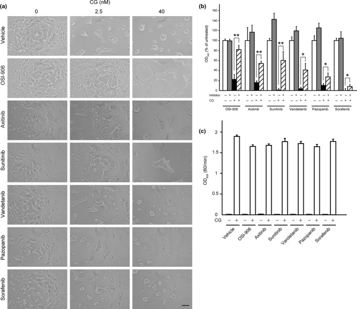
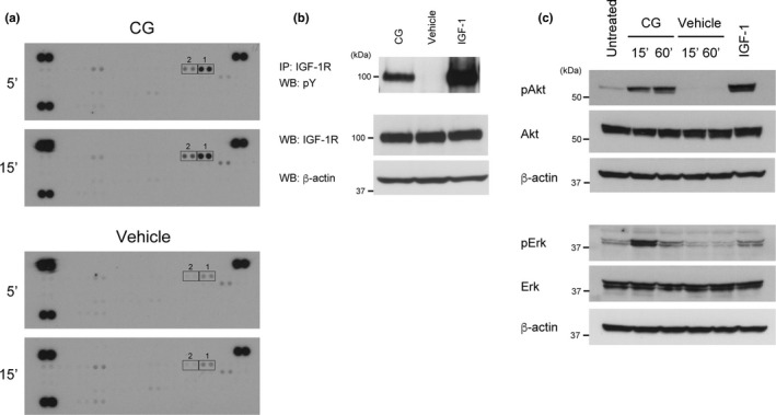
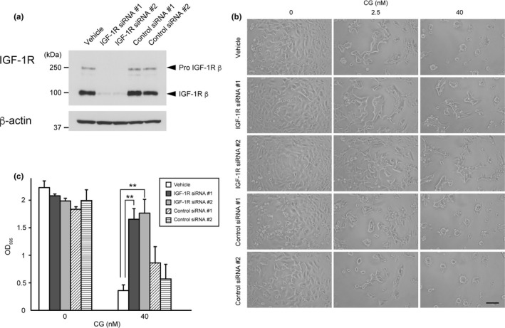
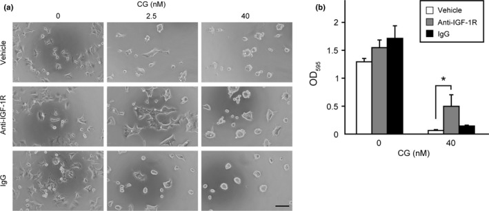
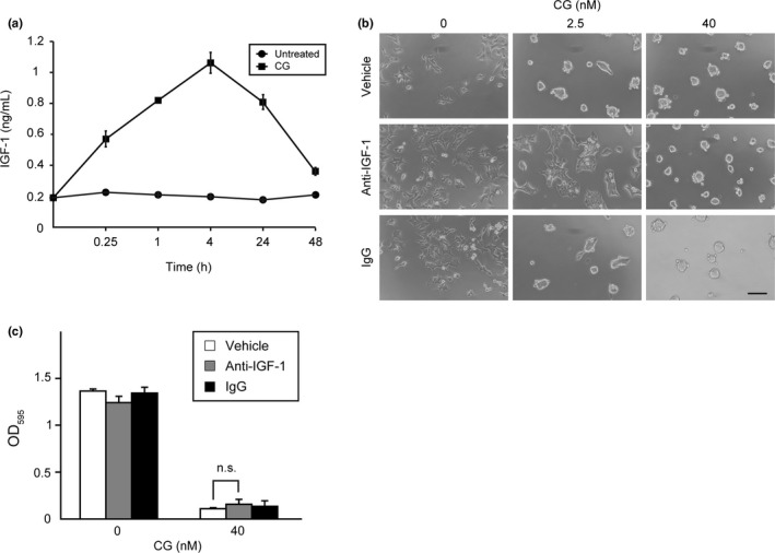
Cathepsin G (CG), a neutrophil serine protease, induces cell migration and multicellular aggregation of human breast cancer MCF-7 cells in a process that is dependent on E-cadherin and CG enzymatic activity. While these tumor cell aggregates can cause tumor emboli that could represent intravascular growth and extravasation into the surrounding tissues, resulting in metastasis, the molecular mechanism underlying this process remains poorly characterized. In this study, we aimed to identify the signaling pathway that is triggered during CG-mediated stimulation of cell aggregation. Screening of a library of compounds containing approximately 90 molecular-targeting drugs revealed that this process was suppressed by the insulin-like growth factor-1 (IGF-1) receptor (IGF-1R)-specific kinase inhibitor OSI-906, as well as the multikinase inhibitors axitinib and sunitinib. Antibody array analysis, which is capable of detecting tyrosine phosphorylation of 49 distinct receptor tyrosine kinases, and the results of immunoprecipitation studies indicated that IGF-1R is phosphorylated in response to CG treatment. Notably, IGF-1R neutralization via treatment with a specific antibody or silencing of IGF-1R expression through siRNA transfection suppressed cell aggregation. Furthermore, CG treatment of MCF-7 cells resulted in increased release of IGF-1 into the medium for 24 h, while antibody-mediated IGF-1 neutralization partially prevented CG-induced cell aggregation. These results demonstrate that autocrine IGF-1 signaling is partly responsible for the cell aggregation induced by CG.

Keywords: breast neoplasm; neoplasm metastasis; neutrophil; serine protease; tumor microenvironment.


Figures

Figure 1
Figure 1
Cathepsin G (CG)‐induced MCF‐7 cell aggregation is suppressed by insulin‐like growth factor‐1 receptor (IGF‐1R)‐specific and multikinase inhibitors. (a) Morphology of cells incubated concurrently for 24 h with CG and kinase inhibitors (OSI‐906, 10 μM; axitinib, 10 μM; sunitinib, 5 μM; vandetanib, 10 μM; pazopanib, 10 μM; sorafenib, 5 μM). Scale bar = 100 μm. (b) Comparison of the inhibitory effects of various tested agents on CG‐induced MCF‐7 cell aggregation. The degree of cell aggregation was quantified using a cell‐aggregation assay. Cells were incubated with CG (40 nM) and respective inhibitors (OSI‐906, 10 μM; axitinib, 10 μM; sunitinib, 5 μM; vandetanib, 10 μM; pazopanib, 10 μM; sorafenib, 5 μM) for 24 h and then washed, following which the residual cells were stained with crystal violet. The results are expressed as means ± SD (n = 3); *P < 0.05, **P < 0.001, Student's t‐test. (c) OSI‐906 and multikinase inhibitors did not affect the enzymatic activity of CG. The enzymatic activity of CG was analyzed by measuring the rate of 4‐nitroanilide release after incubation of N‐succinyl‐Ala‐Ala‐Pro‐Phe p‐nitroanilide with CG (40 nM) and the respective inhibitors (OSI‐906, 10 μM; axitinib, 10 μM; sunitinib, 5 μM; vandetanib, 5 μM; erlotinib, 10 μM; pazopanib, 10 μM; sorafenib, 5 μM). The results are expressed as means ± SD (n = 3).
Figure 2
Figure 2
Cathepsin G (CG) induces the phosphorylation of insulin‐like growth factor‐1 receptor (IGF‐1R) and insulin receptor. (a) CG‐stimulated phosphorylation of receptor tyrosine kinases in the Proteome Profiler Human Phospho‐RTK array system. Cell lysates (300 μg/membrane) were prepared from cells treated with CG (40 nM) for 5 or 15 min. The spots were blotted with antibodies against receptor tyrosine kinases, and all antibodies were spotted on the membranes in duplicate (Spots 1: IGF‐1R, spots 2: insulin receptor). (b) Tyrosine phosphorylation of IGF‐1R stimulated by CG was detected by immunoprecipitation using an anti‐IGF‐1R antibody and western blotting with an anti‐phosphotyrosine antibody. (c) CG evokes Akt and Erk phosphorylation. Cell lysates (20 μg) prepared from cells incubated with CG (40 nM) for the indicated time periods were subjected to western blot analysis.
Figure 3
Figure 3
Downregulation of insulin‐like growth factor‐1 receptor (IGF‐1R) expression attenuates cathepsin G (CG)‐induced cell aggregation. (a) Western blot analysis of IGF‐1R protein expression within the lysates (20 μg) of cells transfected with IGF‐1R‐specific siRNA using an anti‐IGF‐1R β‐subunit antibody. (b) Morphology of IGF‐1R siRNA‐expressing cells following incubation with CG for 24 h. Scale bar = 100 μm. (c) Effect of IGF‐1R knockdown on CG‐induced MCF‐7 cell aggregation. The degree of cell aggregation was quantified using a cell‐aggregation assay. After washing cells that had been incubated with CG (40 nM) for 24 h, the residual cells were stained with crystal violet. The results are expressed as means ± SD (n = 3); **P < 0.001, Student's t test.
Figure 4
Figure 4
Antibody‐mediated insulin‐like growth factor‐1 receptor (IGF‐1R) neutralization attenuates cathepsin G (CG)‐induced cell aggregation. (a) Morphology of MCF‐7 cells incubated concurrently with CG and an anti‐IGF‐1R α‐subunit antibody (10 μg/mL) for 24 h. Scale bar = 100 μm. (b) Effect of an anti‐IGF‐1R α‐subunit antibody on CG‐induced MCF‐7 cell aggregation. The degree of cell aggregation was quantified using a cell‐aggregation assay. Cells were incubated with CG (40 nM) for 24 h and then washed, following which the residual cells were stained with crystal violet. The results are expressed as means ± SD (n = 3); *P < 0.05, Student's t‐test.
Figure 5
Figure 5
Insulin‐like growth factor‐1 (IGF‐1) released from cathepsin G (CG)‐treated MCF‐7 cells causes cell aggregation. (a) Graphic depiction of the amounts of free IGF‐1 in conditioned medium obtained from MCF‐7 cells stimulated with CG (40 nM). The results are presented as means ± SD (n = 3). (b) IGF‐1 neutralization using a specific antibody attenuates CG‐induced cell aggregation. MCF‐7 cells were incubated with CG and an anti‐IGF‐1 antibody (10 μg/mL) for 24 h. Scale bar = 100 μm. (c) Quantitative analysis of the effect of an anti‐IGF‐1 antibody on CG‐induced MCF‐7 cell aggregation. The degree of cell aggregation was quantified using a cell‐aggregation assay. After washing the cells that had been incubated with CG (40 nM) for 24 h, the residual cells were stained with crystal violet. The results are expressed as means ± SD (n = 3); n.s., not significant, Student's t test.

Similar articles

Cited by

References

    1. Coussens LM, Werb Z. Inflammation and cancer. Nature 2002; 420: 860–7. - PMC - PubMed
    1. Elinav E, Nowarski R, Thaiss CA, Hu B, Jin C, Flavell RA. Inflammation‐induced cancer: crosstalk between tumours, immune cells and microorganisms. Nat Rev Cancer 2013; 13: 759–71. - PubMed
    1. Fridlender ZG, Sun J, Kim S et al Polarization of tumor‐associated neutrophil phenotype by TGF‐β: “N1” versus “N2” TAN. Cancer Cell 2009; 16: 183–94. - PMC - PubMed
    1. Gregory AD, Houghton AM. Tumor‐associated neutrophils: new targets for cancer therapy. Cancer Res 2011; 71: 2411–6. - PubMed
    1. Fridlender ZG, Albelda SM. Tumor‐associated neutrophils: friend or foe? Carcinogenesis 2012; 33: 949–55. - PubMed

MeSH terms